...
For convenience you can obtain the publications specifically based on TCIA in Endnote XML format: Pubs_basedon_TCIA_0617TCIA0218.xml. This This should be usable as input to your favorite reference management system.
...
TCIA-Related Publication History
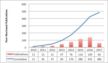
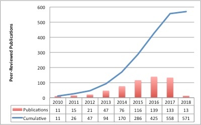
Table of Contents
| Table of Contents | ||||
|---|---|---|---|---|
|
TCIA General
- PriorGillmann, FC., SmithArbelaez, KP., SharmaPenaloza, A., Kirby, J. , Tarbox, LT., ClarkHagen, KH., Bennett, W., Nolan, T., Freymann, J& Wischgoll, T. (2017). The public cancer radiology imaging collections of The Cancer Imaging Archive. Nature Scientific Data, 4; 1-7. Intuitive error space exploration of medical image data in clinical daily routine. Eurographics Conference on Visualization (EuroVis) 2017. doi: 10.10382312/eurovisshort.20171148
- Jinu, J., Rajesh, K. R., Pournami, S. C., & Vidya, Psdata.2017.124 Kohli, M., Morrison, J. J., Wawira, J., Morgan, M. B., & Hostetter, J., Genereaux, B., Hussain, M., Langer S. G. (2017). Creation and curation of the society of imaging informatics in medicine hackathon dataset. Journal of Digital Imaging, 1-4Interactive 3D Virtual Colonoscopic Navigation For Polyp Detection From CT Images. Procedia Computer Science, 115, 407-414. doi: 10.1007/s10278-017-0003-51016/j.procs.2017.09.099
- Ghosh, D., & Bandyopadhyay, S. KWilliamson, J.F., Das, S.K., Goodsitt, M.S., Deasy, J.O. (2017). Introducing the Medical Physics Dataset Article. Med. Phys. 44(2); 349-350. doi: 10.1002/mp.12003
- Nida, N; Khan, M. (2016). Efficient Colorization of Medical Imaging based on Colour Transfer Method. U.G. Proceedings of the Pakistan Academy of Sciences: B. Life and Environmental Sciences, 53(4); 253-261. (link)
- Kalpathy-Cramer, J., Zhao, B., Goldgof, D., Gu, Y., Wang, X., Yang, H., Tan, Y., Gillies, R., Napel, S. (2016). A Comparison of Lung Nodule Segmentation Algorithms: Methods and Results from a Multi-institutional Study. J Digit Imaging 29(4):476-487. DOI: 10.1007/s10278-016-9859-z
Parks, C.L., Monson, K.L. (2016). Automated Facial Recognition of Computed Tomography-Derived Facial Images: Patient Privacy Implications. Journal of Digital Imaging. 1-11. DOI: 10.1007/s10278-016-9932-7
Huang, B.E., Mulyasasmita, W., Rajagopal, G. (2016). The Path from Big Data to Precision Medicine. Expert Review of Precision Medicine and Drug Development, 1(2):129-143. (link)
- Chatellier, G., Varlet, V., Blachier-Poisson, C. (2016). "Big data" and "open data": What kind of access should researchers enjoy? Therapie. 71(1); 97-105, 107-114.(link)
- Benedict, S.H., Hoffman K., Martel, M.K., Abernethy, A.P., Asher, A.L., Capala, J., Chen, R.C., Chera, B., Couch, J., Deye, J., Efstathiou, J.A., Ford, E., Fraass, B.A., Gabriel, P.E., Huser, V., Kavanagh, B.D., Khuntia, D., Marks, L.B., Mayo, C., McNutt, T., Miller, R.S., Moore, K.L., Prior, F., Roelofs, E., Rosenstein, B.S., Sloan, J., Theriault, A., Vikram, B. (2016). Overview of the American Society for Radiation Oncology–National Institutes of Health–American Association of Physicists in Medicine Workshop 2015: Exploring Opportunities for Radiation Oncology in the Era of Big Data. International Journal of Radiation Oncology: Biology, Physics. 95(3):873-879 (link)
- Toga, A.W., Dinov, I.D. (2015). Sharing big biomedical data. Journal of Big Data. 2(1); 1-12. (link)
- Moore, S.M., Maffitt, D.R., Smith, K.E., Kirby, J.S., Clark, K.W., Freymann, J.B., Vendt, B.A., Tarbox, L.R., Prior, F.W. (2015). De-identification of Medical Images with Retention of Scientific Research Value. RadioGraphics. 35(3); 727-35. doi: 10.1148/rg.2015140244.
- Mayo, C.S., Deasy, J.O., Chera, B.S., Freymann, J., Kirby, J.S., Hardenberg, P.H. (2016). How Can We Effect Culture Change Toward Data-Driven Medicine? International Journal of Radiation Oncology: Biology, Physics. 95(3); 916-21. (link)
- Kirby, J., Tarbox, L., Freymann, J., Jaffe, C., Prior, F. (2015). "TU-AB-BRA-03: The Cancer Imaging Archive: Supporting Radiomic and Imaging Genomic Research with Open-Access Data Sets." Medical physics 42(6): 3587-3587. DOI: 10.1118/1.4925508
- GIllies, R.J., Kinahan, P.E., Hricak, H., (2016). Radiomics: Images Are More than Pictures, They Are Data. Radiology, 278(2); 563-77. (link)
- Fedorov, A., Clunie, D., Ulrich, E., Bauer, C., Wahle, A., Brown, B., Onken, M., Riesmeier, J., Pieper, S., Kikinis, R., Buatti, J., Beichel, R.R. (2016). DICOM for quantitative imaging biomarker development: A standards based approach to sharing of clinical data and structured PET/CT analysis results in head and neck cancer research. PeerJ, 4(e2057). (link)
- Commean, P.K., Rathmell, J.M., Clark, K.W., Maffitt, D.R., Prior, F.W. (2015). A Query Tool for Investigator Access to the Data and Images of the National Lung Screening Trial. Journal of Digital Imaging. 1-9. (paper)
- Bourne, P.E. (2015). DOIs for DICOM Raw Images: Enabling Science Reproducibility. Radiology. 275(1); 3-4. link.
- Armato, S.G., Hadjiiski, L., Tourassi, G.D., Drukker, K., Giger, M.L., Li, F., Redmond, G., Farahani, K., Kirby, J.S., Clarke, L.P. (2015). Special Section Guest Editorial: LUNGx Challenge for computerized lung nodule classification: reflections and lessons learned. Journal of Medical Imaging. 2(2); 10.1117/1.JMI.2.2.020103
- Herskovits, E.H. (2014). Quantitative Radiology: Applications to Oncology. Emerging Applications of Molecular Imaging to Oncology. 124; 1-30. (link)
- Gutman, D.A., Dunn Jr., W.D., Cobb, J., Stoner, R.M., Kalpathy-Cramer, J., Erickson, B. (2014) Web based tools for visualizing imaging data and development of XNATView, a zero footprint image viewer. Frontiers in Neuroinformatics. 8. (paper)
- Erickson, B.J., Fajnwaks, P., Langer, S.G., and Perry, J. (2014) Multisite Image Data Collection and Management Using the RSNA Image Sharing Network., Translational oncology, 7(1); 36-39. (paper)
- Prior, F.W., Clark, K., Commean, P., Freymann, J., Jaffe, C., Kirby, J., Moore, S., Smith, K., Tarbox, L., Vendt, B. (2013) TCIA: an information resource to enable open science. Engineering in Medicine and Biology Society (EMBC), 2013 35th Annual International Conference of the IEEE. (paper)
- Gutman, D.A., Cobb, J., Somanna, D., Park, Y., Wang, F., Kurc, T., Saltz, J.H., Brat, D.J., Cooper, L.A. (2013) Cancer Digital Slide Archive: an informatics resource to support integrated in silico analysis of TCGA pathology data. Journal of the American Medical Informatics Association. 20(6); 1091-1098. doi: 10.1136/amiajnl-2012-001469 (paper)
- Clark, K., Vendt, B., Smith, K., Freymann, J., Kirby, J., Koppel, P., Moore, S., Phillips, S., Maffitt, D., Pringle, M., Tarbox, L., Prior, F. (2013). The Cancer Imaging Archive (TCIA): Maintaining and Operating a Public Information Repository, Journal of Digital Imaging, 26(6), 1045-1057. (paper)
- Villani, L., and Prati, R.C. (2012). Classificação Multirrótulo na Anotação Automática de Nódulo Pulmonar Solitário. Congresso Brasileiro de Informática em Saúde, Citado na. (paper)
- Mongkolwat, P., Channin, D.S., Kleper, V., Rubin, D.L. (2012). Informatics in Radiology: An Open-Source and Open-Access Cancer Biomedical Informatics Grid Annotation and Image Markup Template Builder. Radiographics, 32(4); 1223-32. (paper).
- Jaffe, C.C. (2012). Imaging and Genomics: Is There a Synergy? Radiology. 264(2); 329-31.(paper).
Freymann, J.B., Kirby, J.S., Perry, J.H., Clunie, D.A., Jaffe, C.C. (2012). Image data sharing for biomedical research—meeting HIPAA requirements for de-identification. Journal of Digital Imaging, 25(1). 14-24. (paper)
Radiogenomics
Lehrer, M., Bhadra, A., Ravikumar, V., Chen, J. Y., Wintermark, M., Hwang, S. N., Holder, C. A., Huang, E. P., Fevrier-Sullivan, B., Freymann, J. B., Rao, A., & TCGA Glioma Phenotype Research Group. (2017). Multiple-response regression analysis links magnetic resonance imaging features to de-regulated protein expression and pathway activity in lower grade glioma. Oncoscience, 4, 57-66. doi:10.18632/oncoscience.353
- Demerath, T., Simon-Gabriel, C.P., Kellner, E., Schwarzwald, R., Lange, T., Heiland, D.H., Reinacher, P., Staszewski, O., Mast, H., Kiselev, V.G., Egger, K., Urbach, H., Weyerbrock, A., Mader, I. (2017). Mesoscopic imaging of glioblastomas: Are diffusion, perfusion and spectroscopic measures influenced by the radiogenetic phenotype? Neuroradiology Journal, 30(1); 36-47. doi: 10.1177/1971400916678225
Liu, T.T., Achrol, A.S., Mitchell, L.A., Rodriguez, S.A., Feroze, A., Iv, M., Kim, C., Chaudhary, N., Gevaert, O., Stuart, J.M., Harsh, G.R., Chang, S.D., Rubin, D.L. (2016). Magnetic resonance perfusion image features uncover an angiogenic subgroup of glioblastoma patients with poor survival and better response to antiangiogenic treatment. Neuro-Oncology, 1-11. doi: 10.1093/neuonc/now270
Schrock, M., Batar, B., Lee, J., Druck, T., Ferguson, B., Cho, J., Akakpo, K., Hagrass, H., Heerema, N., Xia, F. (2016). Wwox–Brca1 interaction: role in DNA repair pathway choice. Oncogene, 1-13. doi: 10.1038/onc.2016.389.
Song, S.E., Bae, M.S., Chang, J.M., Cho, N., Ryu, H.S., Moon, W.K. (2016). MR and mammographic imaging features of HER2-positive breast cancers according to hormone receptor status: a retrospective comparative study. Acta Radiologica. 58(7), 792-799. doi: 10.1177/0284185116673119
McCann, S.M., Jiang, Y., Fan, X., Wang, J. Antic, T., Prior, F., VanderWeele, D., Oto, A. Quantitative Multiparametric MRI Features and PTEN Expression of Peripheral Zone Prostate Cancer: A Pilot Study. American Journal of Roentgenology 206(3); 559-565 doi: 10.2214/AJR.15.14967
Katrib, A., Hsu, W., Bui, A., Xing, Y. (2016). “Radiotranscriptomics”: A synergy of imaging and transcriptomics in clinical assessment. Quantitative Biology. 1-12. doi: 10.1007/s40484-016-0061-6
- Bai, H.X., Lee, A.M., Yang, L., Zhang, P., Davatzikos, C., Maris, J.M., Diskin, S.J. (2016). Imaging genomics in cancer research: Limitations and promises. The British Journal of Radiology, 89(1061); doi: 10.1259/bjr.20151030
Zhu, Y., H. Li, et al. (2015). TU-CD-BRB-06: Deciphering Genomic Underpinnings of Quantitative MRI-Based Radiomic Phenotypes of Invasive Breast Carcinoma. Medical physics 42(6): 3603-3603. doi: 10.1118/1.4925591
Tomczak, K., Czerwińska, P., Wiznerowicz, M. (2015). The Cancer Genome Atlas (TCGA): an immeasurable source of knowledge. Contemp Oncol (Pozn). 19(1A); A68-A77. doi: 10.5114/wo.2014.47136
- Shinegare, A.B., Vikram, R., Jaffe, C., Akin, O., Kirby, J., Huang, E., Freymann, J., Sainani, N.I., Sadow, C.A., Bathala, T.K., Rubin, D.L., Oto, A., Heller, M.T., Surabhi, V.R., Katabathina, V., Silverman, S.G. (2015). Radiogenomics of clear renal cell carcinoma: Preliminary Findings of The Cancer Genome Atlas-Renal Cell Carcinoma (TCGA-RCC) Imaging Research Group. Abdominal Imaging, 40(6). 1684-1692. doi: 10.1007/s00261-015-0386-z
Pope, W.B. (2015). Genomics of Brain Tumor Imaging. Neuroimaging Clinics of North America. 25(1); 105-19. doi: 10.1016/j.nic.2014.09.006
- Gutman, D.A., Dunn Jr., W.D., Grossmann, P., Cooper, L.A., Holder, C.A., Ligon, K.L., Alexander, B.M., Aerts, H.J. (2015). Somatic mutations associated with MRI-derived volumetric features in glioblastoma. Neuroradiology, 57(12); 1227-1237. doi: 10.1007/s00234-015-1576-7
Feldman, M., Piazza, M.G., Edwards, N.A., Ray, Chaudhury, A., Maric, D., Merrill, M.J., Zhuang, Z., Chittiboina, P. (2015). 137 Somatostatin Receptor Expression on VHL-Associated Hemangioblastomas Offers Novel Therapeutic Target. Neurosurgery 62. (CN_suppl_1); 209-210. doi: 10.1227/01.neu.0000467099.84064.25
- Colen R, Foster I, Gatenby R, Giger ME, Gillies R, Gutman D, Heller M, Jain R, Madabhushi A, Madhavan S, Napel S, Rao A, Saltz J, Tatum J, Verhaak R, Whitman G. (2014). NCI Workshop Report: Clinical and Computational Requirements for Correlating Imaging Phenotypes with Genomics Signatures. Translational Oncology. 7(5); 556-69. doi: 10.1016/j.tranon.2014.07.007.
- Rao A. (2013). Exploring relationships between multivariate radiological phenotypes and genetic features: A case-study in Glioblastoma using the Cancer Genome Atlas, Global Conference on Signal and Information Processing (GlobalSIP), doi: 10.1109/GlobalSIP.2013.6736815
Gevaert, O., Xu, J., Hoang, C.D., Leung, A.N., Xu, Y., Quon, A., Rubin, D.L., Napel, S., Plevritis, S.K. (2012) Non-small cell lung cancer: identifying prognostic imaging biomarkers by leveraging public gene expression microarray data--methods and preliminary results. Radiology. 264(2); 387-96. doi: 10.1148/radiol.12111607
Radiomics
Vani, N., Swomya, A., & Jayamma, N. (2017). MRI Brain tumor classification using support vector machine. International Research Journal of Engineering and Technology, 1724-1729. doi: 10.1109/SCEECS.2014.6804439
- Beichel, R.R., Smith, B.J., Bauer, C., Ulrich, E.J., Ahmadvand, P., Budzevich, M.M., Gillies, R.J., Goldgof, D., Grkovski, M., Hamarneh, G., Huang, Q., Kinahan, P.E., Laymon, C.M., Mountz, J.M., Muzi, J.P., Muzi, M., Nehmeh, S., Oborski, M.J., Tan, Y., Zhao, B., Sunderland, J.J., Buatti, J.M. (2017). Multi-site quality and variability analysis of 3D FDG PET segmentations based on phantom and clinical image data. Med. Phys. 44(2); 479-496. doi: 10.1002/mp.12041
- Vallières, M., Kay-Rivest, E., Perrin, L.J., Liem, X., Furstoss, C., Aerts, H.J.W.L., Khaouam, N., Nguyen-Tan, P.F., Want, C.-S., Sultanem, K., Seuntjens, J., Naqa, I.E. (2017). Radiomics strategies for risk assessment of tumour failure in head-and-neck cancer. Scientific Reports, (arXiv 1703.08516)
- Paredes, D., Saha, A., Mazurowski, M.A.(2017). Deep learning for segmentation of brain tumors: can we train with images from different institutions? SPIE Medical Imaging: Computer-Aided Diagnosis, doi: 10.1117/12.2255696
- Kumar, S., Dharun. (2017). Combination of fuzzy c-means clustering and texture pattern matrix for brain MRI segmentation. Biomedical Research, 28(5)
- Nabizadeh N, Kubat M. Automatic Tumor Segmentation in Single-Spectral MRI Using A Texture-Based and Contour-Based Algorithm. ScienceDirect, 77: 1-10. doi: 10.1016/j.eswa.2017.01.036
Kaur, T., Saini, B.S., Gupta, S. (2016). A joint intensity and edge magnitude-based multilevel thresholding algorithm for the automatic segmentation of pathological MR brain images. Neural Computing and Applications. 1-24. doi: 10.1007/s00521-016-2751-4
Song, J., Liu, Z., Zhong, W., Huang, Y., Ma, Z., Dong, D., Liang, C., Tian, J. (2016). Non-small cell lung cancer: quantitative phenotypic analysis of CT images as a potential marker of prognosis. Scientific Reports. 6:38282:1-9. doi: 10.1038/srep38282
Crawford, L., Monod, A., Chen, A.X., Mukherjee, S., Rabadán, R. (2016). Topological Summaries of Tumor Images Improve Prediction of Disease Free Survival in Glioblastoma Multiforme. arXiv preprint arXiv:161106818.
- Korfiatis, P., Kline, T.L., Erickson, B.J. (2016). Automated Segmentation of Hyperintense Regions in FLAIR MRI Using Deep Learning. J Tomography, 2(4) 334-340 DOI: 10.18383/j.tom.2016.00166
Zheng, C., Wang, X., Feng, D. (Eds.). (2016). Topology guided demons registration with local rigidity preservation. 2016 IEEE 38th Annual International Conference Engineering in Medicine and Biology Society (EMBC). IEEE. doi: 10.1109/EMBC.2016.7590913
Kotrotsou, A., Zinn, P.O., Colen, R.R. (2016). Radiomics in Brain Tumors: An Emerging Technique for Characterization of Tumor Environment. Magnetic Resonance Imaging Clinics of North America. 24(4); 719-29. doi: 10.1016/j.mric.2016.06.006
- Zhao, B., Tan, Y., Tsai, W.Y., Qi, J., Xie, C., Lu, L., Schwartz, L.H. (2016). Reproducibility of radiomics for deciphering tumor phenotype with imaging. Scientific Reports. 6:23428. doi: 10.1038/srep23428
- Li, H., Zhu, Y., Burnside, E.S., Huang, E., Drukker, K., Hoadley, K.A., Fan, C., Conzen, S.D., Zuley, M., Net, J.M., Sutton, E., Whitman, G.J., Morris, E., Perou, C.M., Ji, Y., Giger, M.L. (2016). Quantitative MRI radiomics in the prediction of molecular classifications of breast cancer subtypes in the TCGA/TCIA data set. npj Breast Cancer. doi: 10.1038/npjbcancer.2016.12
- Grossmann, P., Gutman, D.A., Dunn Jr., W.D., Holder, C.A., Aerts, H.J.W.L. (2016). Imaging-genomics reveals driving pathways of MRI derived volumetric tumor phenotype features in Glioblastoma. BMC Cancer. 16(611). doi: 10.1186/s12885-016-2659-5
- Zhu, Y., Li, H., Guo, W., Drukker, K., Lian, L., Giger, M.L., Ji, Y. (2015). Deciphering Genomic Underpinnings of Quantitative MRI-based Radiomic Phenotypes of Invasive Breast Carcinoma. Scientific Reports. 5(17787). doi: 10.1038/srep17787
- Rajakumar, K., Muttan, S., Deepa, G., Revathy, S., Priya, B.S. (2015). Intelligent texture feature extraction and indexing for MRI image retrieval using curvelet and PCA with HTF. Advances in Natural and Applied Sciences. 9(6 SE) 506-513. doi: (link)
Parmar, C., Leijenaar, R.T.H., Grossmann, P., Valazquez, E.R., Bussink, J., Rietveld, D., Rietbergen, M.M., Haibe-Kains, B., Lambin, P., Aerts, H.J.W.L. (2015). Radiomic feature clusters and Prognostic Signatures specific for Lung and Head &Neck cancer. Scientific Reports. 5(11044) doi: 10.1038/srep11044
- Parmar, C., Grossmann, P., Bussink, J., Lambin, P., Aerts, H.J.W.L. (2015). Machine Learning methods for Quantitative Radiomic Biomarkers. Scientific Reports, 5(13087). doi: 10.1038/srep13087
- Chaddad, A., Tanougast, C. (2015), High-Throughput Quantification of Phenotype Heterogeneity Using Statistical Features. Advances in Bioinformatics, 15(728164). doi: 10.1155/2015/728164
- Chaddad, A. (2015). Automated Feature Extraction in Brain Tumor by Magnetic Resonance Imaging Using Gaussian Mixture Models. International Journal of Biomedical Imaging, 2015(868031). doi: 10.1155/2015/868031
- Dhara, A.K., Mukhopadhyay, S., Khandelwal, N. (2013). 3d texture analysis of solitary pulmonary nodules using co-occurrence matrix from volumetric lung CT images. Medical Imaging 2013: Computer-Aided Diagnosis, 8670. doi: 10.1117/12.2007016
Dhara, A.K., Mukhopadhyay, S., Alam, N., Khandelwal, N. (2013). Measurement of spiculation index in 3D for solitary pulmonary nodules in volumetric lung CT images.Medical Imaging 2013: Computer-Aided Diagnosis, 8670. doi: 10.1117/12.2006970
Algorithm Development
- Brain tumor detection from MRI image: An approach. International Journal of Applied Research, 3(6), 1152-1159. Retrieved from https://pdfs.semanticscholar.org/1916/f00997b627213b46c874a9a133ee8b6fa92e.pdf
- Vallières, M., Laberge, S., Diamant, A., & El Naqa, I. (2017). Enhancement of multimodality texture-based prediction models via optimization of PET and MR image acquisition protocols: a proof of concept. Physics in Medicine and Biology, 62(22), 8536-8565.
- Mitra, S., Banerjee, S., & Hayashi, Y. (2017). Volumetric brain tumour detection from MRI using visual saliency. (J. Najbauer, Ed.) PLOS One, 12(11). http://dx.doi.org/10.1371/journal.pone.0187209
Gueziri, H.-E. (2017). User-centered design and evaluation of interactive segmentation methods for medical images. Montreal: École de technologie supérieure du Quebec. Retrieved from http://espace.etsmtl.ca/1959/2/GUEZIRI_Houssem-Eddine-web.pdf
Lan, R., Zhong, S., Liu, Z., Shi, Z., & Luo, X. (2017). A simple texture feature for retrieval of medical images. Multimedia Tools and Applications. DOI: 10.1007/s11042-017-5341-2
Prior, F., Smith, K., Sharma, A., Kirby, J., Tarbox, L., Clark, K., Bennett, W., Nolan, T., Freymann, J. (2017). The public cancer radiology imaging collections of The Cancer Imaging Archive. Nature Scientific Data, 4; 1-7. doi:10.1038/sdata.2017.124
Kohli, M., Morrison, J. J., Wawira, J., Morgan, M. B., & Hostetter, J., Genereaux, B., Hussain, M., Langer S. G. (2017). Creation and curation of the society of imaging informatics in medicine hackathon dataset. Journal of Digital Imaging, 1-4. doi:10.1007/s10278-017-0003-5
- Williamson, J.F., Das, S.K., Goodsitt, M.S., Deasy, J.O. (2017). Introducing the Medical Physics Dataset Article. Med. Phys. 44(2); 349-350. doi: 10.1002/mp.12003
- Nida, N; Khan, M. (2016). Efficient Colorization of Medical Imaging based on Colour Transfer Method. U.G. Proceedings of the Pakistan Academy of Sciences: B. Life and Environmental Sciences, 53(4); 253-261. (link)
- Kalpathy-Cramer, J., Zhao, B., Goldgof, D., Gu, Y., Wang, X., Yang, H., Tan, Y., Gillies, R., Napel, S. (2016). A Comparison of Lung Nodule Segmentation Algorithms: Methods and Results from a Multi-institutional Study. J Digit Imaging29(4):476-487. DOI: 10.1007/s10278-016-9859-z
Parks, C.L., Monson, K.L. (2016). Automated Facial Recognition of Computed Tomography-Derived Facial Images: Patient Privacy Implications. Journal of Digital Imaging. 1-11. DOI: 10.1007/s10278-016-9932-7
Huang, B.E., Mulyasasmita, W., Rajagopal, G. (2016). The Path from Big Data to Precision Medicine.Expert Review of Precision Medicine and Drug Development,1(2):129-143. (link)
- Chatellier, G., Varlet, V., Blachier-Poisson, C. (2016). "Big data" and "open data": What kind of access should researchers enjoy?Therapie. 71(1); 97-105, 107-114.(link)
- Benedict, S.H., Hoffman K., Martel, M.K., Abernethy, A.P., Asher, A.L., Capala, J., Chen, R.C., Chera, B., Couch, J., Deye, J., Efstathiou, J.A., Ford, E., Fraass, B.A., Gabriel, P.E., Huser, V., Kavanagh, B.D., Khuntia, D., Marks, L.B., Mayo, C., McNutt, T., Miller, R.S., Moore, K.L., Prior, F., Roelofs, E., Rosenstein, B.S., Sloan, J., Theriault, A., Vikram, B. (2016). Overview of the American Society for Radiation Oncology–National Institutes of Health–American Association of Physicists in Medicine Workshop 2015: Exploring Opportunities for Radiation Oncology in the Era of Big Data.International Journal of Radiation Oncology: Biology, Physics. 95(3):873-879 (link)
- Toga, A.W., Dinov, I.D. (2015). Sharing big biomedical data.Journal of Big Data. 2(1); 1-12. (link)
- Moore, S.M., Maffitt, D.R., Smith, K.E., Kirby, J.S., Clark, K.W., Freymann, J.B., Vendt, B.A., Tarbox, L.R., Prior, F.W. (2015). De-identification of Medical Images with Retention of Scientific Research Value. RadioGraphics. 35(3); 727-35. doi: 10.1148/rg.2015140244.
- Mayo, C.S., Deasy, J.O., Chera, B.S., Freymann, J., Kirby, J.S., Hardenberg, P.H. (2016). How Can We Effect Culture Change Toward Data-Driven Medicine?International Journal of Radiation Oncology: Biology, Physics. 95(3); 916-21. (link)
- Kirby, J., Tarbox, L., Freymann, J., Jaffe, C., Prior, F. (2015). "TU-AB-BRA-03: The Cancer Imaging Archive: Supporting Radiomic and Imaging Genomic Research with Open-Access Data Sets."Medical physics 42(6): 3587-3587. DOI: 10.1118/1.4925508
- GIllies, R.J., Kinahan, P.E., Hricak, H., (2016). Radiomics: Images Are More than Pictures, They Are Data.Radiology, 278(2); 563-77. (link)
- Fedorov, A., Clunie, D., Ulrich, E., Bauer, C., Wahle, A., Brown, B., Onken, M., Riesmeier, J., Pieper, S., Kikinis, R., Buatti, J., Beichel, R.R. (2016). DICOM for quantitative imaging biomarker development: A standards based approach to sharing of clinical data and structured PET/CT analysis results in head and neck cancer research.PeerJ, 4(e2057). (link)
- Commean, P.K., Rathmell, J.M., Clark, K.W., Maffitt, D.R., Prior, F.W. (2015). A Query Tool for Investigator Access to the Data and Images of the National Lung Screening Trial. Journal of Digital Imaging. 1-9. (paper)
- Bourne, P.E. (2015). DOIs for DICOM Raw Images: Enabling Science Reproducibility. Radiology. 275(1); 3-4. link.
- Armato, S.G., Hadjiiski, L., Tourassi, G.D., Drukker, K., Giger, M.L., Li, F., Redmond, G., Farahani, K., Kirby, J.S., Clarke, L.P. (2015). Special Section Guest Editorial: LUNGx Challenge for computerized lung nodule classification: reflections and lessons learned. Journal of Medical Imaging. 2(2); 10.1117/1.JMI.2.2.020103
- Herskovits, E.H. (2014). Quantitative Radiology: Applications to Oncology. Emerging Applications of Molecular Imaging to Oncology. 124; 1-30. (link)
- Gutman, D.A., Dunn Jr., W.D., Cobb, J., Stoner, R.M., Kalpathy-Cramer, J., Erickson, B. (2014) Web based tools for visualizing imaging data and development of XNATView, a zero footprint image viewer. Frontiers in Neuroinformatics. 8. (paper)
- Erickson, B.J., Fajnwaks, P., Langer, S.G., and Perry, J. (2014) Multisite Image Data Collection and Management Using the RSNA Image Sharing Network., Translational oncology, 7(1); 36-39. (paper)
- Prior, F.W., Clark, K., Commean, P., Freymann, J., Jaffe, C., Kirby, J., Moore, S., Smith, K., Tarbox, L., Vendt, B. (2013) TCIA: an information resource to enable open science. Engineering in Medicine and Biology Society (EMBC), 2013 35th Annual International Conference of the IEEE.(paper)
- Gutman, D.A., Cobb, J., Somanna, D., Park, Y., Wang, F., Kurc, T., Saltz, J.H., Brat, D.J., Cooper, L.A. (2013) Cancer Digital Slide Archive: an informatics resource to support integrated in silico analysis of TCGA pathology data.Journal of the American Medical Informatics Association. 20(6); 1091-1098. doi: 10.1136/amiajnl-2012-001469 (paper)
- Clark, K., Vendt, B., Smith, K., Freymann, J., Kirby, J., Koppel, P., Moore, S., Phillips, S., Maffitt, D., Pringle, M., Tarbox, L., Prior, F. (2013). The Cancer Imaging Archive (TCIA): Maintaining and Operating a Public Information Repository, Journal of Digital Imaging, 26(6), 1045-1057. (paper)
- Villani, L., and Prati, R.C. (2012). Classificação Multirrótulo na Anotação Automática de Nódulo Pulmonar Solitário.Congresso Brasileiro de Informática em Saúde, Citado na. (paper)
- Mongkolwat, P., Channin, D.S., Kleper, V., Rubin, D.L. (2012). Informatics in Radiology: An Open-Source and Open-Access Cancer Biomedical Informatics Grid Annotation and Image Markup Template Builder. Radiographics, 32(4); 1223-32. (paper).
- Jaffe, C.C. (2012). Imaging and Genomics: Is There a Synergy?Radiology. 264(2); 329-31.(paper).
Freymann, J.B., Kirby, J.S., Perry, J.H., Clunie, D.A., Jaffe, C.C. (2012). Image data sharing for biomedical research—meeting HIPAA requirements for de-identification. Journal of Digital Imaging, 25(1). 14-24. (paper)
Radiogenomics
- Lee, J., Cui, Y., Sun, X., Li, B., Wu, J., Li, D., Gensheimer, M. F., Loo Jr., B. W., Diehn, M., Li, R. (2017). Prognostic value and molecular correlates of a CT image-based quantitative pleural contact index in early stage NSCLC. European Radiology, 1-11. Retrieved from https://link.springer.com/article/10.1007/s00330-017-4996-4. DOI: 10.1007/s00330-017-4996-4
Smits, M., & van den Bent, M. J. (2017). Imaging correlates of adult glioma genotypes. Radiology, 284(2). http://dx.doi.org/10.1148/radiol.2017151930
Lehrer, M., Bhadra, A., Ravikumar, V., Chen, J. Y., Wintermark, M., Hwang, S. N., Holder, C. A., Huang, E. P., Fevrier-Sullivan, B., Freymann, J. B., Rao, A., & TCGA Glioma Phenotype Research Group. (2017). Multiple-response regression analysis links magnetic resonance imaging features to de-regulated protein expression and pathway activity in lower grade glioma. Oncoscience, 4, 57-66. doi:10.18632/oncoscience.353
- Demerath, T., Simon-Gabriel, C.P., Kellner, E., Schwarzwald, R., Lange, T., Heiland, D.H., Reinacher, P., Staszewski, O., Mast, H., Kiselev, V.G., Egger, K., Urbach, H., Weyerbrock, A., Mader, I. (2017). Mesoscopic imaging of glioblastomas: Are diffusion, perfusion and spectroscopic measures influenced by the radiogenetic phenotype? Neuroradiology Journal, 30(1); 36-47. doi: 10.1177/1971400916678225
Liu, T.T., Achrol, A.S., Mitchell, L.A., Rodriguez, S.A., Feroze, A., Iv, M., Kim, C., Chaudhary, N., Gevaert, O., Stuart, J.M., Harsh, G.R., Chang, S.D., Rubin, D.L. (2016). Magnetic resonance perfusion image features uncover an angiogenic subgroup of glioblastoma patients with poor survival and better response to antiangiogenic treatment. Neuro-Oncology, 1-11. doi: 10.1093/neuonc/now270
Schrock, M., Batar, B., Lee, J., Druck, T., Ferguson, B., Cho, J., Akakpo, K., Hagrass, H., Heerema, N., Xia, F. (2016). Wwox–Brca1 interaction: role in DNA repair pathway choice. Oncogene, 1-13. doi: 10.1038/onc.2016.389.
Song, S.E., Bae, M.S., Chang, J.M., Cho, N., Ryu, H.S., Moon, W.K. (2016). MR and mammographic imaging features of HER2-positive breast cancers according to hormone receptor status: a retrospective comparative study. Acta Radiologica. 58(7), 792-799. doi: 10.1177/0284185116673119
McCann, S.M., Jiang, Y., Fan, X., Wang, J. Antic, T., Prior, F., VanderWeele, D., Oto, A. Quantitative Multiparametric MRI Features and PTEN Expression of Peripheral Zone Prostate Cancer: A Pilot Study. American Journal of Roentgenology 206(3); 559-565 doi: 10.2214/AJR.15.14967
Katrib, A., Hsu, W., Bui, A., Xing, Y. (2016). “Radiotranscriptomics”: A synergy of imaging and transcriptomics in clinical assessment.Quantitative Biology. 1-12. doi: 10.1007/s40484-016-0061-6
- Bai, H.X., Lee, A.M., Yang, L., Zhang, P., Davatzikos, C., Maris, J.M., Diskin, S.J. (2016). Imaging genomics in cancer research: Limitations and promises.The British Journal of Radiology, 89(1061); doi: 10.1259/bjr.20151030
Zhu, Y., H. Li, et al. (2015). TU-CD-BRB-06: Deciphering Genomic Underpinnings of Quantitative MRI-Based Radiomic Phenotypes of Invasive Breast Carcinoma.Medical physics 42(6): 3603-3603. doi: 10.1118/1.4925591
Tomczak, K., Czerwińska, P., Wiznerowicz, M. (2015). The Cancer Genome Atlas (TCGA): an immeasurable source of knowledge.Contemp Oncol (Pozn). 19(1A); A68-A77. doi: 10.5114/wo.2014.47136
- Shinegare, A.B., Vikram, R., Jaffe, C., Akin, O., Kirby, J., Huang, E., Freymann, J., Sainani, N.I., Sadow, C.A., Bathala, T.K., Rubin, D.L., Oto, A., Heller, M.T., Surabhi, V.R., Katabathina, V., Silverman, S.G. (2015). Radiogenomics of clear renal cell carcinoma: Preliminary Findings of The Cancer Genome Atlas-Renal Cell Carcinoma (TCGA-RCC) Imaging Research Group.Abdominal Imaging, 40(6). 1684-1692. doi: 10.1007/s00261-015-0386-z
Pope, W.B. (2015). Genomics of Brain Tumor Imaging. Neuroimaging Clinics of North America. 25(1); 105-19. doi: 10.1016/j.nic.2014.09.006
- Gutman, D.A., Dunn Jr., W.D., Grossmann, P., Cooper, L.A., Holder, C.A., Ligon, K.L., Alexander, B.M., Aerts, H.J. (2015). Somatic mutations associated with MRI-derived volumetric features in glioblastoma.Neuroradiology, 57(12); 1227-1237. doi: 10.1007/s00234-015-1576-7
Feldman, M., Piazza, M.G., Edwards, N.A., Ray, Chaudhury, A., Maric, D., Merrill, M.J., Zhuang, Z., Chittiboina, P. (2015). 137 Somatostatin Receptor Expression on VHL-Associated Hemangioblastomas Offers Novel Therapeutic Target.Neurosurgery 62. (CN_suppl_1); 209-210. doi: 10.1227/01.neu.0000467099.84064.25
- Colen R, Foster I, Gatenby R, Giger ME, Gillies R, Gutman D, Heller M, Jain R, Madabhushi A, Madhavan S, Napel S, Rao A, Saltz J, Tatum J, Verhaak R, Whitman G. (2014). NCI Workshop Report: Clinical and Computational Requirements for Correlating Imaging Phenotypes with Genomics Signatures. Translational Oncology. 7(5); 556-69. doi: 10.1016/j.tranon.2014.07.007.
- Rao A. (2013). Exploring relationships between multivariate radiological phenotypes and genetic features: A case-study in Glioblastoma using the Cancer Genome Atlas, Global Conference on Signal and Information Processing (GlobalSIP), doi: 10.1109/GlobalSIP.2013.6736815
Gevaert, O., Xu, J., Hoang, C.D., Leung, A.N., Xu, Y., Quon, A., Rubin, D.L., Napel, S., Plevritis, S.K. (2012) Non-small cell lung cancer: identifying prognostic imaging biomarkers by leveraging public gene expression microarray data--methods and preliminary results. Radiology. 264(2); 387-96. doi: 10.1148/radiol.12111607
Radiomics
Larue, R. T. H. M., Van De Voorde, L., van Timmeren, J. E., Leijenaar, Ralph T. H., Berbee, M., Sosef, M. N., Schreurs, W. M. J., van Elmpt, W., Lambin, P. (2017). 4DCT imaging to assess radiomics feature stability: An investigation for thoracic cancers. Radiotherapy and Oncology. DOI: 10.1016/j.radonc.2017.07.023
Sutton, E. J., Huang, E. P., Drukker, K., Burnside, E. S., Li, H., Net, J. M., Rao, A., Whitman, G. J., Zuley, M., Ganott, M., Bonaccio, E., Giger, M. L., Morris, E. A. (2017). Breast MRI radiomics: Comparison of computer- and human-extracted imaging phenotypes. European Radiology Experimental. http://dx.doi.org/10.1186/s41747-017-0025-2
Vani, N., Swomya, A., & Jayamma, N. (2017). MRI Brain tumor classification using support vector machine. International Research Journal of Engineering and Technology, 1724-1729. doi: 10.1109/SCEECS.2014.6804439
- Beichel, R.R., Smith, B.J., Bauer, C., Ulrich, E.J., Ahmadvand, P., Budzevich, M.M., Gillies, R.J., Goldgof, D., Grkovski, M., Hamarneh, G., Huang, Q., Kinahan, P.E., Laymon, C.M., Mountz, J.M., Muzi, J.P., Muzi, M., Nehmeh, S., Oborski, M.J., Tan, Y., Zhao, B., Sunderland, J.J., Buatti, J.M. (2017). Multi-site quality and variability analysis of 3D FDG PET segmentations based on phantom and clinical image data. Med. Phys. 44(2); 479-496. doi: 10.1002/mp.12041
- Vallières, M., Kay-Rivest, E., Perrin, L.J., Liem, X., Furstoss, C., Aerts, H.J.W.L., Khaouam, N., Nguyen-Tan, P.F., Want, C.-S., Sultanem, K., Seuntjens, J., Naqa, I.E. (2017). Radiomics strategies for risk assessment of tumour failure in head-and-neck cancer. Scientific Reports, (arXiv 1703.08516)
- Paredes, D., Saha, A., Mazurowski, M.A.(2017). Deep learning for segmentation of brain tumors: can we train with images from different institutions?SPIE Medical Imaging: Computer-Aided Diagnosis, doi: 10.1117/12.2255696
- Kumar, S., Dharun. (2017). Combination of fuzzy c-means clustering and texture pattern matrix for brain MRI segmentation. Biomedical Research, 28(5)
- Nabizadeh N, Kubat M. Automatic Tumor Segmentation in Single-Spectral MRI Using A Texture-Based and Contour-Based Algorithm. ScienceDirect, 77: 1-10. doi: 10.1016/j.eswa.2017.01.036
Kaur, T., Saini, B.S., Gupta, S. (2016). A joint intensity and edge magnitude-based multilevel thresholding algorithm for the automatic segmentation of pathological MR brain images. Neural Computing and Applications. 1-24. doi: 10.1007/s00521-016-2751-4
Song, J., Liu, Z., Zhong, W., Huang, Y., Ma, Z., Dong, D., Liang, C., Tian, J. (2016). Non-small cell lung cancer: quantitative phenotypic analysis of CT images as a potential marker of prognosis. Scientific Reports. 6:38282:1-9. doi: 10.1038/srep38282
Crawford, L., Monod, A., Chen, A.X., Mukherjee, S., Rabadán, R. (2016). Topological Summaries of Tumor Images Improve Prediction of Disease Free Survival in Glioblastoma Multiforme. arXiv preprint arXiv:161106818.
- Korfiatis, P., Kline, T.L., Erickson, B.J. (2016). Automated Segmentation of Hyperintense Regions in FLAIR MRI Using Deep Learning. J Tomography, 2(4) 334-340 DOI: 10.18383/j.tom.2016.00166
Zheng, C., Wang, X., Feng, D. (Eds.). (2016). Topology guided demons registration with local rigidity preservation. 2016 IEEE 38th Annual International Conference Engineering in Medicine and Biology Society (EMBC). IEEE. doi: 10.1109/EMBC.2016.7590913
Kotrotsou, A., Zinn, P.O., Colen, R.R. (2016). Radiomics in Brain Tumors: An Emerging Technique for Characterization of Tumor Environment. Magnetic Resonance Imaging Clinics of North America. 24(4); 719-29. doi: 10.1016/j.mric.2016.06.006
- Zhao, B., Tan, Y., Tsai, W.Y., Qi, J., Xie, C., Lu, L., Schwartz, L.H. (2016). Reproducibility of radiomics for deciphering tumor phenotype with imaging. Scientific Reports. 6:23428. doi: 10.1038/srep23428
- Li, H., Zhu, Y., Burnside, E.S., Huang, E., Drukker, K., Hoadley, K.A., Fan, C., Conzen, S.D., Zuley, M., Net, J.M., Sutton, E., Whitman, G.J., Morris, E., Perou, C.M., Ji, Y., Giger, M.L. (2016). Quantitative MRI radiomics in the prediction of molecular classifications of breast cancer subtypes in the TCGA/TCIA data set. npj Breast Cancer. doi: 10.1038/npjbcancer.2016.12
- Grossmann, P., Gutman, D.A., Dunn Jr., W.D., Holder, C.A., Aerts, H.J.W.L. (2016). Imaging-genomics reveals driving pathways of MRI derived volumetric tumor phenotype features in Glioblastoma. BMC Cancer. 16(611). doi: 10.1186/s12885-016-2659-5
- Zhu, Y., Li, H., Guo, W., Drukker, K., Lian, L., Giger, M.L., Ji, Y. (2015). Deciphering Genomic Underpinnings of Quantitative MRI-based Radiomic Phenotypes of Invasive Breast Carcinoma. Scientific Reports. 5(17787). doi: 10.1038/srep17787
- Rajakumar, K., Muttan, S., Deepa, G., Revathy, S., Priya, B.S. (2015). Intelligent texture feature extraction and indexing for MRI image retrieval using curvelet and PCA with HTF. Advances in Natural and Applied Sciences. 9(6 SE) 506-513. doi: (link)
Parmar, C., Leijenaar, R.T.H., Grossmann, P., Valazquez, E.R., Bussink, J., Rietveld, D., Rietbergen, M.M., Haibe-Kains, B., Lambin, P., Aerts, H.J.W.L. (2015). Radiomic feature clusters and Prognostic Signatures specific for Lung and Head &Neck cancer.Scientific Reports. 5(11044) doi: 10.1038/srep11044
- Parmar, C., Grossmann, P., Bussink, J., Lambin, P., Aerts, H.J.W.L. (2015). Machine Learning methods for Quantitative Radiomic Biomarkers. Scientific Reports, 5(13087). doi: 10.1038/srep13087
- Chaddad, A., Tanougast, C. (2015), High-Throughput Quantification of Phenotype Heterogeneity Using Statistical Features. Advances in Bioinformatics, 15(728164). doi: 10.1155/2015/728164
- Chaddad, A. (2015). Automated Feature Extraction in Brain Tumor by Magnetic Resonance Imaging Using Gaussian Mixture Models. International Journal of Biomedical Imaging, 2015(868031). doi: 10.1155/2015/868031
- Dhara, A.K., Mukhopadhyay, S., Khandelwal, N. (2013). 3d texture analysis of solitary pulmonary nodules using co-occurrence matrix from volumetric lung CT images. Medical Imaging 2013: Computer-Aided Diagnosis, 8670. doi: 10.1117/12.2007016
Dhara, A.K., Mukhopadhyay, S., Alam, N., Khandelwal, N. (2013). Measurement of spiculation index in 3D for solitary pulmonary nodules in volumetric lung CT images.Medical Imaging 2013: Computer-Aided Diagnosis, 8670. doi: 10.1117/12.2006970
Algorithm Development
- Babu, J. S., Mathew, S., & Simon, R. (2017). Biomedical image retrieval using LBWP. International Research Journal of Engineering and Technology (IRJET), 4(9), 839-843. https://www.irjet.net/archives/V4/i9/IRJET-V4I9147.pdf
Hostetter, J. M., Morrison, J. J., Morris, M., Jeudy, J., Wang, K. C., & Siegel, E. (2017). Personalizing lung cancer risk prediction and imaging follow-up recommendations using the National Lung Screening Trial dataset. Journal of the American Medical Informatics Association, 24(6), 1046-1051. doi:10.1093/jamia/ocx012
- Mason J, Perelli A, Nailon W, Davies M. Can Planning Images Reduce Scatter in Follow-Up Cone-Beam CT?arXiv 1703.07179 2017.
Hsieh KL-C, Tsai R-J, Teng Y-C, Lo C-M. Effect of a computer-aided diagnosis system on radiologists' performance in grading gliomas with MRI. PloS one. 2017;12(2):e0171342 (link)
Hsieh KL-C, Lo C-M, Hsiao C-J. Computer-aided grading of gliomas based on local and global MRI features. Computer Methods and Programs in Biomedicine. 2017;139:31-8. DOI: doi.org/10.1016/
- Mason J, Perelli A, Nailon W, Davies M. Can Planning Images Reduce Scatter in Follow-Up Cone-Beam CT? arXiv 1703.07179 2017.
Hsieh KL-C, Tsai R-J, Teng Y-C, Lo C-M. Effect of a computer-aided diagnosis system on radiologists' performance in grading gliomas with MRI. PloS one. 2017;12(2):e0171342 (link)
Hsieh KL-C, Lo C-M, Hsiao C-J. Computer-aided grading of gliomas based on local and global MRI features. Computer Methods and Programs in Biomedicine. 2017;139:31-8. DOI: doi.org/10.1016/j.cmpb.2016.10.021
Yang H, Liu F, Wang Z, Tang H, Sun S, Sun S. Research on the Content-Based Classification of Medical Image. Journal of Medical Imaging and Health Informatics. 2017;7(1):129-36. (link)
Rezaie AA, Habiboghli A. Detection of Lung Nodules on Medical Images by the Use of Fractal Segmentation. International Journal of Interactive Multimedia and Artificial Inteligence. 2017;4(Special Issue on 3D Medicine and Artificial Intelligence):15-9. (link)
Chen H, Zhang Y, Zhang W, Liao P, Li K, Zhou J, Wang G. Low-dose CT via convolutional neural network. Biomedical Optics Express. 2017;8(2):679-94.(link)
- Vallières M, Freeman C, Skamene S, El Naqa I. A radiomics model from joint FDG-PET and MRI texture features for the prediction of lung metastases in soft-tissue sarcomas of the extremities.Physics in medicine and biology. 2015;60(14):5471.
- Kazdal S, Dogan B, Camurcu AY, editors. Computer-aided detection of brain tumors using image processing techniques. Signal Processing and Communications Applications Conference (SIU), 2015 23th; 2015: IEEE.
- Gupta A, Martens O, Le Moullec Y, Saar T, editors. A tool for lung nodules analysis based on segmentation and morphological operation. Intelligent Signal Processing (WISP), 2015 IEEE 9th International Symposium on; 2015: IEEE.
- Benninghoff H, Garcke H. Segmentation of Three-dimensional Images with Parametric Active Surfaces and Topology Changes. arXiv:1506.07136. 2015.
- Zabala-Travers S, Choi M, Cheng W-C, Badano A. Effect of color visualization and display hardware on the visual assessment of pseudocolor medical images. Medical Physics. 2015;42(6):2942-54.
- Guvenis A, Koc A. OPTIMISING DELINEATION ACCURACY OF TUMOURS IN PET FOR RADIOTHERAPY PLANNING USING BLIND DECONVOLUTION. Radiation Protection Dosimetry. 2015:ncv110.
- Grove O, Berglund AE, Schabath MB, Aerts HJ, Dekker A, Wang H, Velazquez ER, Lambin P, Gu Y, Balagurunathan Y. Quantitative Computed Tomographic Descriptors Associate Tumor Shape Complexity and Intratumor Heterogeneity with Prognosis in Lung Adenocarcinoma. PloS one. 2015;10(3).
- Buerger C, Sénégas J, Kabus S, Carolus H, Schulz H, Agarwal H, Turkbey B, Choyke P, Renisch S. Comparing nonrigid registration techniques for motion corrected MR prostate diffusion imaging. Medical physics. 2015;42(1):69-80.
- Abedini M, Codella N, Connell J, Garnavi R, Merler M, Pankanti S, Smith J, Syeda-Mahmood T. A generalized framework for medical image classification and recognition. IBM Journal of Research and Development. 2015;59(2/3):1: -: 18.
- Blessy SPS, Sulochana CH. Performance analysis of unsupervised optimal fuzzy clustering algorithm for MRI brain tumor segmentation. Technology and Health Care. 2014.
- ElNawasany AM, Ali AF, Waheed ME. A Novel Hybrid Perceptron Neural Network Algorithm for Classifying Breast MRI Tumors. Advanced Machine Learning Technologies and Applications: Springer; 2014. p. 357-66.
- Hong S, Huang Y, Cao Y, Chen X, Han J-DJ. Approaches to uncovering cancer diagnostic and prognostic molecular signatures. Molecular & Cellular Oncology. 2014.
- Codella N, Connell J, Pankanti S, Merler M, and Smith JR. Automated Medical Image Modality Recognition by Fusion of Visual and Text Information. Medical Image Computing and Computer-Assisted Intervention. 2014, Springer. 487-495. (link)
- Ertugrul OF. Adaptive Texture Energy Measure Method. International Journal of Intelligent Information Systems. 2014. 3(2):13-18. doi:10.11648/j.ijiis.20140302.11 (link)
- Kawa J, Juszczyk J, Pyciński B, Badura P, Pietka E. Radiological Atlas for Patient Specific Model Generation. Information Technologies in Biomedicine, 2014 4:69-82. 10.1007/978-3-319-06596-0_7. (link)
- Kowalik-Urbaniak I, Brunet D, Wang J, Koff D, Smolarski-Koff N, Vrscay ER, Wallace B, Wang Z.The quest for ‘diagnostically lossless’ medical image compression: a comparative study of objective quality metrics for compressed medical images. SPIE Medical Imaging. 2014. Vol. 9073. International Society for Optics and Photonics. doi:10.1117/12.2043196 (link)
- Naresh P and Shettar R. Image Processing and Classification Techniques for Early Detection of Lung Cancer for Preventive Health Care: A Survey. International Journal of Recent Trends in Engineering & Technology, 2014. 11:595-601 (link)
- Patel NP, Parmar SK, and Jain KR. Swift Pre Rendering Volumetric Visualization of Magnetic Resonance Cardiac Images based on Isosurface Technique. Procedia Technology, 2014. 14:422-429. doi:10.1016/j.protcy.2014.08.054 (link)
- Roy S, Brown MS, and Shih GL. Visual Interpretation with Three-Dimensional Annotations (VITA): Three-Dimensional Image Interpretation Tool for Radiological Reporting. Journal of Digital Imaging, 2014. 27(1):49-57. doi: 10.1007/s10278-013-9624-5 (link)
Roth HR, Lu L, Seff A, Cherry KM, Hoffman J, Wang S, Liu J, Turkbey E, Summers RM. A new 2.5 D representation for lymph node detection using random sets of deep convolutional neural network observations. Medical Image Computing and Computer-Assisted Intervention–MICCAI 2014: Springer; 2014. p. 520-7.
- Sivakumar S, and Chandrasekar C. A Study on Image Denoising for Lung CT Scan Images.International Journal of Emerging Technologies in Computational and Applied Sciences, 2014. 7(1):86-91 (link)
Seff A, Lu L, Cherry KM, Roth HR, Liu J, Wang S, Hoffman J, Turkbey EB, Summers RM. 2d view aggregation for lymph node detection using a shallow hierarchy of linear classifiers. Medical Image Computing and Computer-Assisted Intervention–MICCAI 2014: Springer; 2014. p. 544-52.
- Harmon S, Wendelberger B, and Jeraj R. SU-E-J-98: Radiogenomics: Correspondence Between Imaging and Genetic Features Based On Clustering Analysis. Medical Physics, 2014. 41(6): p. 178-178. doi:10.1118/1.4888150 (link)
- Krishnakumar V. and Parthiban L. Performance Analysis of Denoising in MR Images with Double Density Dual Tree Complex Wavelets, Curvelets and NonSubsampled Contourlet Transforms. Annual Review & Research in Biology, 2014. 4(19):2938-2956. doi:10.9734/ARRB/2014/9131#sthash.qFePVdL1.dpuf (link)
- Codella N, Merler M. IBM TJ Watson Research Center. Semantic Model Vector for ImageCLEF2013. June 18, 2014. (link)
- Agostinelli F, Anderson MR, and Lee H. Adaptive Multi-Column Deep Neural Networks with Application to Robust Image Denoising. Advances in Neural Information Processing Systems. 2013. (link)
Agostinelli F, Anderson MR, Lee H, editors. Robust Image Denoising with Multi-Column Deep Neural Networks. Advances in Neural Information Processing Systems; 2013.
- Breseman K, Lee C, Bloch BN, and Jaffe C. Constructing 3D-Printable CAD Models of Prostates from MR Images. Bioengineering Conference (NEBEC),
39th Annual Northeast , IEEE, 27-28. 5-7 April 2013. doi:10.1109/NEBEC.2013.8 - Buckler A, Liu TT, Savig E, Suzek BE, Rubin DL, and Paik D. Quantitative Imaging Biomarker Ontology (QIBO) for Knowledge Representation of Biomedical Imaging Biomarkers. Journal of Digital Imaging, 2013. 26(4):630-641. doi:10.1007/s10278-013-9599-2 (link)
- Heyns M, Breseman K, Lee C, Bloch BN, Jaffe C, and Xiang H. Design of a Patient-Specific Radiotherapy Treatment Target. Bioengineering Conference (NEBEC), 2013 39th Annual Northeast. 2013.171-172. IEEE.doi:10.1109/NEBEC.2013.75
- Kumar A, Kim J, Cai W, Fulham M, and Feng D. Content-Based Medical Image Retrieval: A Survey of Applications to Multidimensional and Multimodality Data. Journal of Digital Imaging, 2013. 26(6):1025-1039. doi: 10.1007/s10278-013-9619-2.(link)
- Lundström C. vPSNR: a visualization-aware image fidelity metric tailored for diagnostic imaging. International Journal of Computer Assisted Radiology and Surgery, 2013. 8(3):437-450. doi: 10.1007/s11548-012-0792-4 (link)
- Olmedo I, Guerra Perez Y, Johnson JF, Raut L, Hoe DHK. Image segmentation on GPGPUs: a cellular automata-based approach. Proceedings of the 2013 Summer Computer Simulation Conference. Society for Modeling & Simulation International. 2013. 51. (link)
- Pambrun JF, Noumeir R. Compressibility variations of JPEG2000 compressed computed tomography. Conference Proceedings, 35th Annual International Conference of the IEEE Engineering in Medicine and Biology Society, 2013:3375-3378. doi: 10.1109/EMBC.2013.6610265 (link)
- Roozgard A, Barzigar N, Verma P, and Cheng S. 3D medical image denoising using 3D block matching and low-rank matrix completion. Signals, Systems and Computers, Asilomar Conference, 3-6 Nov. 2013, 253 – 257 IEEE. doi:10.1109/ACSSC.2013.6810271
- Yankeelov TE, Atuegwu N, Hormuth D, et al. Clinically Relevant Modeling of Tumor Growth and Treatment Response. Sci Transl Med. 2013 May 29;5(187):187ps9 doi: 10.1126/scitranslmed.3005686 (link) .
- Huang LC, Yseng LY, Hwang MS. A reversible data hiding method by histogram shifting in high quality medical images. Journal of Systems and Software 2013 March;86(3):716-27 doi: 10.1016/j.jss.2012.11.024 (link)
- Pheng HS and Shamsuddin SM. Texture classification of lung computed tomography images. 2012 International Conference on Graphic and Image Processing. 2013. Vol. 8768. International Society for Optics and Photonics. doi:10.1117/12.2011108 (link)
- Barzigar N, Roozgard A, Verma P, Cheng S. Removing Mixture Noise from Medical Images Using Block Matching Filtering and Low-Rank Matrix Completion. Healthcare Informatics, Imaging and Systems Biology, IEEE International Conference. 2012.134. doi:10.1109/HISB.2012.59 (link)
- Otake Y, Schafer S, Stayman JW, Zbijewski W, Kleinszig G, Graumann R, Khanna AJ, Siewerdsen JH. Automatic localization of target vertebrae in spine surgery using fast CT-to-fluoroscopy (3D-2D) image registration. SPIE Medical Imaging, 2012. Volume: 8316. International Society for Optics and Photonics. doi:10.1117/12.911308 (link)
- Roozgard A, Cheng AS, Liu H. Malignant nodule detection on lung ct scan images with kernel rx-algorithm. Biomedical and Health Informatics (BHI), 2012 IEEE-EMBS International Conference on 5-7 Jan. 2012 499 – 502. IEEE. doi: 10.1109/BHI.2012.6211627.
- Biancardi AM, Jirapatnakul AC, Reeves AP. A comparison of ground truth estimation methods. International Journal of Computer Assisted Radiology and Surgery, 2010. 5(3):295-305. doi: 10.1007/s11548-009-0401-3 (link)
- Soysal OM, Chen P, Schneider H. An Image Processing Tool for Efficient Feature Extraction in Computer-Aided Detection Systems. Granular Computing (GrC) IEEE International Conference 2010. 14-16 Aug. 438-442. doi:10.1109/GrC.2010.128
- Tseng LY and Huang LC. Automatic fissure detection in CT images based on the genetic algorithm. Machine Learning and Cybernetics (ICMLC), International Conference. IEEE. 2010. 5: 2583 – 2588. doi: 10.1109/ICMLC.2010.5580871
Kumar, D., A. Wong, et al. (2015). Lung Nodule Classification Using Deep Features in CT Images. Computer and Robot Vision (CRV), 2015 12th Conference on, IEEE.
Kanas, V. G., E. I. Zacharaki, et al. (2015). "A low cost approach for brain tumor segmentation based on intensity modeling and 3D Random Walker." Biomedical Signal Processing and Control 22: 19-30.
Magdy, E., N. Zayed, et al. (2015). "Automatic Classification of Normal and Cancer Lung CT Images Using Multiscale AM-FM Features." International Journal of Biomedical Imaging 2015.
Zayed, N. and H. A. Elnemr (2015). "Statistical Analysis of Haralick Texture Features to Discriminate Lung Abnormalities." International Journal of Biomedical Imaging 2015.
- Chaddad, A. and C. Tanougast "High-Throughput Quantification of Phenotype Heterogeneity Using Statistical Features." Advances in Bioinformatics 2015. doi: 10.1155/2015/728164
- Li M, Miller K, Joldes GR, Kikinis R, Wittek A. Biomechanical model for computing deformations for whole-body image registration: A meshless approach. InternationalJournal for Numerical Methods in Biomedical Engineering. 2016. doi: 10.1002/cnm.2771
...
Androutsou, T. Clinical Decision Support System for Lung Cancer Diagnosis by analysis of thoracic CT images. Carrier NTUA, Department of Electrical and Computer Engineering 2017. (link to thesis)
- Emirzade, Erkan. A COMPUTER AIDED DIAGNOSIS SYSTEM FOR LUNG CANCER DETECTION USING SVM. The Graduate School Of Applied Sciences Of Near East University, 2016. (link to thesis)
- Yu, Zexi. Co-Segmentation Methods for Improving Tumor Target Delineation in PET-CT Images. University of Saskatchewan 2016. (link to thesis)
Albalooshi FA. Self-organizing Approach to Learn a Level-set Function for Object Segmentation in Complex Background Environments. University of Dayton; 2015. (link to thesis)
Camlica Z. Image Area Reduction for Efficient Medical Image Retrieval. Waterloo, Ontario, Canada,: University of Waterloo; 2015. (link to thesis)
- Hunter L. Radiomics of NSCLC: Quantitative CT Image Feature Characterization and Tumor Shrinkage Prediction. Thesis, University of Texas; 2013. (link to thesis)
Karnayana PM. Radiogenomic correlation for prognosis in patients with glioblastoma multiformae.
University of Saskatchewan 2016San Diego State University; 2013. (link to thesis)Albalooshi FA. Self-organizing Approach to Learn a Level-set Function for Object Segmentation in Complex Background Environments. University of Dayton; 2015.
Nabizadeh, N. Automated Brain Lesion Detection and Segmentation Using Magnetic Resonance Images. Electrical and Computer Engineering. Miami, FL, University of Miami. PhD., 2015. (link to thesis)Camlica Z. Image Area Reduction for Efficient Medical Image Retrieval
. Waterloo, Ontario, Canada,: University of Waterloo; 2015Wieser, H.-P. Supervised Machine Learning Approach Utilizing Artificial Neural Networks for Automated Prostate Zone Segmentation in Abdominal MR images. Klagenfurt, Austria, Fachhochschule Kärnten/Carinthia University of Applied Sciences; 2013.(link to thesis)
- Hunter L. Radiomics of NSCLC: Quantitative CT Image Feature Characterization and Tumor Shrinkage Prediction. Thesis, University of Texas; 2013. (link to thesis)
Karnayana PM. Radiogenomic correlation for prognosis in patients with glioblastoma multiformae. San Diego State University; 2013. (link to thesis)
Nabizadeh, N. Automated Brain Lesion Detection and Segmentation Using Magnetic Resonance Images. Electrical and Computer Engineering. Miami, FL, University of Miami. PhD., 2015. (link to thesis)
Wieser, H.-P. Supervised Machine Learning Approach Utilizing Artificial Neural Networks for Automated Prostate Zone Segmentation in Abdominal MR images. Klagenfurt, Austria, Fachhochschule Kärnten/Carinthia University of Applied Sciences; 2013.(link to thesis)
TCIA DOI
TCIA DOI
- Aerts HJ, Velazquez ER, et al. (2014). Decoding tumour phenotype by noninvasive imaging using a quantitative radiomics approach. TCIA. Saint Louis, MO. (link)
- Armato SG and Drukker K, et al. (2015). SPIE-AAPM-NCI Lung Nodule Classification Challenge Dataset. TCIA. Saint Louis, MO. (link)
- Bloch N, Rusu M, et al. (2015) NCI-ISBI 2013 Challenge: Automated Segmentation of Prostate Structures. TCIA. St. Louis, MO. (link)
- Colen RR, Wang J, et al. (2014). Glioblastoma: Imaging Genomic Mapping Reveals Sex-specific Oncogenic Associations of Cell Death. TCIA. Saint Louis, MO. (link)
Gevaert O, Mitchell LA
Aerts HJ, Velazquez ER, et al. (2014).
Decoding tumour phenotype by noninvasive imaging using a quantitative radiomics approachGlioblastoma multiforme: exploratory radiogenomic analysis by using quantitative image features. TCIA. Saint Louis, MO. (link)
- Armato SG and Drukker KGevaert O, Xu J, et al. (20152014). SPIE-AAPM-NCI Lung Nodule Classification Challenge Dataset Non-small cell lung cancer: identifying prognostic imaging biomarkers by leveraging public gene expression microarray data--methods and preliminary results. TCIA. Saint Louis, MO. (link)Bloch
- N, Rusu MGrove O, Berglund AE, et al. (2015) NCI-ISBI 2013 Challenge: Automated Segmentation of Prostate Structures. TCIA. St. Louis, MO. . Data from: Quantitative computed tomographic descriptors associate tumor shape complexity and intratumor heterogeneity with prognosis in lung adenocarcinoma. TCIA. Saint Louis. MO. (link)
- Colen RR, Wang J
Gutman DA, Cooper LA, et al. (2014).
Glioblastoma: Imaging Genomic Mapping Reveals Sex-specific Oncogenic Associations of Cell DeathMR Imaging Predictors of Molecular Profile and Survival: Multi-institutional Study of the TCGA Glioblastoma Data Set. TCIA. Saint Louis, MO. (link)
Gevaert O, Mitchell LAHuang W, Li X, et al. (2014). Glioblastoma multiforme: exploratory radiogenomic analysis by using quantitative image featuresVariations of dynamic contrast-enhanced magnetic resonance imaging in evaluation of breast cancer therapy response: a multicenter data analysis challenge. TCIA. Saint Louis, MO. (link)
Gevaert O, Xu JJain R, Poisson LM, et al. (2014).
Non-small cell lung cancer: identifying prognostic imaging biomarkers by leveraging public gene expression microarray data--methods and preliminary resultsOutcome Prediction in Patients with Glioblastoma by Using Imaging, Clinical, and Genomic Biomarkers: Focus on the Nonenhancing Component of the Tumor. TCIA. Saint Louis, MO. (link)
- Grove O, Berglund AE
Kalpathy-Cramer J, Napel S, et al. (2015).
Data from: Quantitative computed tomographic descriptors associate tumor shape complexity and intratumor heterogeneity with prognosis in lung adenocarcinomaQIN multi-site collection of Lung CT data with Nodule Segmentations. TCIA. Saint Louis
., MO. (link)Gutman DA, Cooper LA
- Lee J, Narang S, et al. (20142015). MR Imaging Predictors of Molecular Profile and Survival: Multi-institutional Study of the TCGA Glioblastoma Data Set Spatial Habitat Features derived from Multiparametric Magnetic Resonance Imaging data from Glioblastoma Multiforme cases. TCIA. Saint Louis, MO. (link)Huang W, Li X
- Liu F, Hernandez-Cabronero M, et al. (20142016). Variations of dynamic contrast-enhanced magnetic resonance imaging in evaluation of breast cancer therapy response: a multicenter data analysis challenge Image Data Used in the Simulations of "The Role of Image Compression Standards in Medical Imaging: Current Status and Future Trends". TCIA. Saint Louis, MO. (link)
- Jain R, Poisson LMMazurowski MA, Zhang J, et al. (2014). Outcome Prediction in Patients with Glioblastoma by Using Imaging, Clinical, and Genomic Biomarkers: Focus on the Nonenhancing Component of the Tumor Radiogenomic Analysis of Breast Cancer: Luminal B Molecular Subtype Is Associated with Enhancement Dynamics at MR Imaging. TCIA. Saint Louis, MO. (link)
Kalpathy-Cramer J, Napel SMessay T, Hardie RC, et al. (20152014). QIN multi-site collection of Lung CT data with Nodule SegmentationsSegmentation of Pulmonary Nodules in Computed Tomography Using a Regression Neural Network Approach and its Application to the Lung Image Database Consortium and Image Database Resource Initiative Dataset. TCIA. Saint Louis, MO. (link)
- Lee J, Narang SMorris E, Burnside M, et al. (2015). Spatial Habitat Features derived from Multiparametric Magnetic Resonance Imaging data from Glioblastoma Multiforme cases2014). TCGA Breast Phenotype Research Group Data sets. TCIA. Saint Louis, MO . (link)
- Liu F, Hernandez-Cabronero M
Roth H, Lu L, et al. (
20162015).
Image Data Used in the Simulations of "The Role of Image Compression Standards in Medical Imaging: Current Status and Future Trends"A new 2.5D representation for lymph node detection in CT. TCIA. Saint Louis, MO. (link)
- Mazurowski MA, Zhang J
Shinagare AB, Vikram R, et al. (
20142015).
Radiogenomic Analysis of Breast Cancer: Luminal B Molecular Subtype Is Associated with Enhancement Dynamics at MR ImagingRadiogenomics of Clear Cell Renal Cell Carcinoma: Preliminary Findings of The Cancer Genome Atlas-Renal Cell Carcinoma (TCGA-RCC) Research Group. TCIA. Saint Louis, MO. (link)
Messay T, Hardie RCVallières M, Freeman CR, et al. (20142015). Segmentation of Pulmonary Nodules in Computed Tomography Using a Regression Neural Network Approach and its Application to the Lung Image Database Consortium and Image Database Resource Initiative DatasetData from: A radiomics model from joint FDG-PET and MRI texture features for the prediction of lung metastases in soft-tissue sarcomas of the extremities. TCIA. Saint Louis, MO. (link)
- Morris E, Burnside M, et al. (2014). TCGA Breast Phenotype Research Group Data sets. TCIA. Saint Louis, MO (link)
Roth H, Lu L, et al. (2015). A new 2.5D representation for lymph node detection in CT. TCIA. Saint Louis, MO. (link)
Shinagare AB, Vikram R, et al. (2015). Radiogenomics of Clear Cell Renal Cell Carcinoma: Preliminary Findings of The Cancer Genome Atlas-Renal Cell Carcinoma (TCGA-RCC) Research Group. TCIA. Saint Louis, MO. (link)
Vallières M, Freeman CR, et al. (2015). Data from: A radiomics model from joint FDG-PET and MRI texture features for the prediction of lung metastases in soft-tissue sarcomas of the extremities. TCIA. Saint Louis, MO. (link)
QIN
- Semmineh NB, Stokes AM, Bell LC, Boxerman JL, Quarles CC. A Population-Based Digital Reference Object (DRO) for Optimizing Dynamic Susceptibility Contrast (DSC)-MRI Methods for Clinical Trials. TOMOGRAPHY, 2017; 3(1)41-9. doi: 10.18383/j.tom.2016.00286
Farahani K, Kalpathy-Cramer J, Chenevert TL, et al. Computational Challenges and Collaborative Projects in the NCI Quantitative Imaging Network. Tomography, 2016;2(4):242-9. (link)
Kalpathy-Cramer J, Mamomov A, Zhao B,et al.. Radiomics of Lung Nodules: A Multi-Institutional Study of Robustness and Agreement of Quantitative Imaging Features. Tomography,2016;2(4):430-7. doi: 10.18383/j.tom.2016.00235.
Huang, W., X. Li, et al. (2014). "Variations of dynamic contrast-enhanced magnetic resonance imaging in evaluation of breast cancer therapy response: a multicenter data analysis challenge." Transl Oncol 7(1): 153-166. (link)
Kalpathy-Cramer J, Freymann JB, Kirby JS, et al. Quantitative Imaging Network: Data Sharing and Competitive Algorithm Validation Leveraging The Cancer Imaging Archive Translational Oncology. 2014 Feb;7(1):147-52. doi: 10.1593/tlo.13862. (link)
- Clarke LP, Nordstrom RJ, Zhang H, Tandon P, et al. The Quantitative Imaging Network: NCI’s Historical Perspective and Planned Goals Translational Oncology. 2014 Feb;7(1):1-4. doi: 10.1593/tlo.13832. (link)
- Levy MA, Freymann JB, Kirby JS, et al. Informatics methods to enable sharing of quantitative imaging research data. Magnetic Resonance Imaging. 2012 Nov;30(9):1249-56. doi: 10.1016/j.mri.2012.04.007. Epub 2012 Jul 6. (link)
Publications relating to specific data collections:
Collection: CT Colonography
Lin AY, Du P, Dinning PG, Arkwright JW, Kamp JP, Cheng LK, Bissett IP, O'Grady G. High resolution anatomic correlation of cyclic motor patterns in the human colon: Evidence of a rectosigmoid brake. American Journal of Physiology-Gastrointestinal and Liver Physiology. 2017;312(5):G508-G15. doi: 10.1152/ajpgi.00021.2017.
Gayathri DK, Radhakrishnan R, Rajamani K. Segmentation of colon and removal of opacified fluid for virtual colonoscopy. Pattern Analysis and Applications. 2017:1-15. doi: 10.1007/s10044-017-0614-y
- Pang S, Yu Z, Orgun MA. A Novel End-to-End Classifier Using Domain Transferred Deep Convolutional Neural Networks for Biomedical Images. Computer Methods and Programs in Biomedicine. 2017. (link)
- Yahya-Zoubir B, Hamami L. et al. Automatic 3D Mesh-Based Centerline Extraction from a Tubular Geometry Form. Information Technology and Control, 2016. 45(2):156-163. (link)
- Alazmani A, Hood A, et al. Quantitative Assessment of Colorectal Morphology: Implications for Robotic Colonoscopy. Medical Engineering and Physics, 2016. 38(2):148-154. (link)
- Gayathri Devi K, Radhakrishnan R. Automatic Segmentation of Colon in 3D CT Images and Removal of Opacified Fluid Using Cascade Feed Forward Neural Network. Computational and Mathematical Methods in Medicine. 2015;2015.
- Namías R, et al., Automatic rectum limit detection by anatomical markers correlation. Computerized Medical Imaging and Graphics, 2014. 38(4):245-250.(link)
- Boone DJ, Halligan S, Roth HR, et al., CT Colonography: External Clinical Validation of an Algorithm for Computer-assisted Prone and Supine Registration. Radiology, 2013. 268(3):752-760.(link)
- Roth HR, et al., External clinical validation of prone and supine CT colonography registration in Abdominal Imaging. Computational and Clinical Applications 2012, Springer. 7601:10-19.(link)
Collection: Head-Neck Cetuximab
Ryalat MH, Laycock S, Fisher M, editors. Automatic Removal of Mechanical Fixations from CT Imagery with Particle Swarm Optimisation. International Conference on Bioinformatics and Biomedical Engineering; 2017: Springer. DOI: 10.1007/978-3-319-56148-6_37
Collection: LIDC-IDRI
- Kang, G., Liu, K., Hou, B., & Zhang, N. (2017). 3D multi-view convolutional neural networks for lung nodule classification. (Y. Deng, Ed.) PLOS One, 12(11). doi: 10.1371/journal.pone.0188290
- Liu, F., Feng, J., Su, W., Lv, Z., Xiao, F., & Qiu, S. (2017). Normalized euclidean super-pixels for medical image segmentation. International Conference on Intelligent Computing (pp. 586-597). Springer. https://link.springer.com/chapter/Ryalat MH, Laycock S, Fisher M, editors. Automatic Removal of Mechanical Fixations from CT Imagery with Particle Swarm Optimisation. International Conference on Bioinformatics and Biomedical Engineering; 2017: Springer. DOI: 10.1007/978-3-319-5614863315-6_37
...
- 2_51
Farag, A. A., Ali, A., Elshazly, S., & Farag, A. A. (2017). Feature fusion for lung nodule classification. International Journal of Computer Assisted Radiology and Surgery, 1-10. doi:10.1007/s11548-017-1626-1
- MC Hancock, JF Magnan. Predictive capabilities of statistical learning methods for lung nodule malignancy classification using diagnostic image features: an investigation using the Lung Image Database Consortium dataset. Proc. SPIE Medical Imaging: Computer-Aided Diagnosis (2017). International Society for Optics and Photonics. doi: 10.1117/12.2254446
- Wang, D; Fong, S; Wong, RK.; Mohammed, S; Fiaidhi, J; Wong, KKL. Robust High-dimensional Bioinformatics Data Streams Mining by ODR-ioVFDT. Scientific Reports 7, article number 43167 doi: 10.1038/srep43167
Mhetre RR, Sache RG. Detection of Lung Cancer Nodule on CT scan Images by using Region Growing Method. International Journal of Current Trends in Engineering & Research. 2016;2(7):215-9. (link)
Setio AAA, Traverso A, de Bel T, Berens MS, Bogaard Cvd, Cerello P, Chen H, Dou Q, Fantacci ME, Geurts B. Validation, comparison, and combination of algorithms for automaticdetection of pulmonary nodules in computed tomography images: the LUNA16 challenge. arXiv preprint arXiv:161208012. 2016:1-16.
- Firmino M, Angelo G, et al. Computer-aided Detection (CADe) and Diagnosis (CADx) System for Lung Cancer with Likelihood of Malignancy Biomed Eng Online (2016) 15(1):2 (link)
- Deep G, Kaur L, et al. Directional Local Ternary Quantized Extrema Pattern: A new descriptor for Biomedical Image Indexing and Retrieval Eng Sci and Tech, an International Journal (2016) (link)
- Wang W, Luo J, Yang X, Lin H. Data Analysis of the Lung Imaging Database Consortium and Image Database Resource Initiative. Academic Radiology. 2015.
Sivakumar, S. and C. Chandrasekar (2015). "A Novel Noise Removal Method for Lung CT SCAN Images Using Statistical Filtering Techniques." International Journal of Algorithms Design and Analysis 1(1).
- Shen S, Bui AA, Cong J, Hsu W. An automated lung segmentation approach using bidirectional chain codes to improve nodule detection accuracy. Computers in biology and medicine. 2015;57:139-49.
- Messay T, Hardie RC, Tuinstra TR. Segmentation of Pulmonary Nodules in Computed Tomography Using a Regression Neural Network Approach and its Application to the Lung Image Database Consortium and Image Database Resource Initiative Dataset. Medical Image Analysis. 2015.(paper)
Magdy, E., N. Zayed, et al. Automatic Classification of Normal and Cancer Lung CT Images using Multi-scale AM-FM Features. Intl Journal of Biomedical Imaging, 2015. (link)
Lassen BC, Jacobs C, et al. Robust Semi-automatic Segmentation of Pulmonary Subsolid Nodules in Chest Computed Tomography Scans. Phys Med Biol (2015) 60(3):1307-1323. (link)
Kumar, D., M. J. Shafiee, et al. Discovery Radiomics for Computed Tomography Cancer Detection. arXiv e-print, 2015. (arXiv link)
Demir, Ö. and A. Yılmaz Çamurcu (2015). "Computer-aided detection of lung nodules using outer surface features." Bio-Medical Materials and Engineering 26(s1): 1213-1222.
Kumar, A., F. Nette, et al. (2014). "A Visual Analytics Approach using the Exploration of Multi-Dimensional Feature Spaces for Content-based Medical Image Retrieval IEEE J Biomed Health Inform (2014) 19(5):1734:1746 (pubmed link)
- Sivakumar S and Chandrasekar C, Lung nodule detection using fuzzy clustering and support vector machines. International Journal of Engineering and Technology, 2013. 5(1):179-185.(link)
- Gavrielides MA, Zeng R, Myers KJ, Sahiner B, Petrick N. Benefit of overlapping reconstruction for improving the quantitative assessment of CT lung nodule volume. Academic Radiology, 2013. 20(2):173-180. doi: 10.1016/j.acra.2012.08.014. (link)
- Aggarwal P, Vig R, and Sardana H Patient-Wise Versus Nodule-Wise Classification of Annotated Pulmonary Nodules using Pathologically Confirmed Cases. Journal of Computers, 2013. 8(9):2245-2255. (link)
- Sivakumar S and Chandrasekar C, Lungs image segmentation through weighted FCM.Recent Advances in Computing and Software Systems (RACSS), 2012 International Conference. 25-27 April 2012 pages 109-113. IEEE. doi:10.1109/RACSS.2012.6212707 (link)
- Armato S, et al., Collaborative projects. Int J CARS, 2012. 7(1):S111-S115.
- Diciotti S, Lombardo S, Falchini M, Picozzi G, Mascalchi M. Automated segmentation refinement of small lung nodules in CT scans by local shape analysis. Biomedical Engineering, IEEE Transactions. 2011. 58(12):3418-3428. doi: 10.1109/TBME.2011.2167621. (link)
Raicu DS, Varutbangkul E, Furst JD, Armato SG III: Modeling semantics from image data: Opportunities from LIDC. International Journal of Biomedical Engineering and Technology 3: 83–113, 2010.
Zinovev D, Duo Y, Raicu DS, Furst JD, Armato SG III: Consensus versus disagreement in imaging research: A case study using the LIDC Database. Journal of Digital Imaging 25: 423–436, 2012.
...
- Stoll M, Stoiber EM, Grimm S, Debus J, Bendl R, Giske K. Comparison of Safety Margin Generation Concepts in Image Guided Radiotherapy to Account for Daily Head and Neck Pose Variations. PloS one. 2016;11(12):e0168916. DOI: 10.1371/journal.pone.0168916
- Ahmadvand P, Duggan N, Bénard F, Hamarneh G. Tumor Lesion Segmentation from 3D PET Using a Machine Learning Driven Active Surface. MLMI 2016 in conjunction with the 19th Int'l Conference on MICCAI. (link)
- Fedorov A, Clunie D, Ulrich E, et al. (2016) DICOM for quantitative imaging biomarker development: a standards based approach to sharing clinical data and structured PET/CT analysis results in head and neck cancer research. PeerJ 4:e2057 DOI: 10.7717/peerj.2057
- Beichel RR, Van Tol M, Ulrich EJ, et al. (2016) Semiautomated segmentation of head and neck cancers in 18F-FDG PET scans: A just-enough-interaction approach. Medical Physics 43:2948–2964. DOI: 10.1118/1.4948679.
Collection: QIN Prostate
- Lavasani, S. N., Mostaar, A., & Ashtiyani, M. (2017). Automatic prostate cancer segmentation using kinetic analysis in dynamic contrast-enhanced MRI. Journal of Biomedical Phsyics and Engineering. doi: 10.22086/jbpe.v0i0.555
- Fedorov A, Fluckiger J, Ayers GD, Li X, Gupta SN, Tempany C, Mulkern R, Yankeelov TE, Fennessy FM. A comparison of two methods for estimating DCE-MRI parameters via individual and cohort based AIFs in prostate cancer: A step towards practical implementation. Magnetic resonance imaging. 2014;32(4):321-9.
- Hegde JV, Mulkern RV, Panych LP, Fennessy FM, Fedorov A, Maier SE, Tempany C. Multiparametric MRI of prostate cancer: An update on state‐of‐the‐art techniques and their performance in detecting and localizing prostate cancer. Journal of Magnetic Resonance Imaging. 2013;37(5):1035-54.
- Benalcázar, M. E., M. Brun, et al. (2015). Automatic Design of Window Operators for the Segmentation of the Prostate Gland in Magnetic Resonance Images. VI Latin American Congress on Biomedical Engineering CLAIB 2014, Paraná, Argentina 29, 30 & 31 October 2014, Springer.
Li, A., C. Li, et al. (2013). Automated Segmentation of Prostate MR Images Using Prior Knowledge Enhanced Random Walker. Digital Image Computing: Techniques and Applications (DICTA), 2013 International Conference on, IEEE.
Qiu, W., J. Yuan, et al. (2014). Prostate segmentation: An efficient convex optimization approach with axial symmetry using 3-D TRUS and MR images. Medical Imaging, IEEE Transactions on 33(4): 947-960.
Xie, Q. and D. Ruan (2014). Low-complexity atlas-based prostate segmentation by combining global, regional, and local metrics. Medical physics 41(4): 041909.
Zhao, T. and D. Ruan (2015). Two-stage fusion set selection in multi-atlas-based image segmentation. Biomedical Imaging (ISBI), 2015 IEEE 12th International Symposium on, IEEE.
...
- Meyer JM, Perlewitz KS, Hayden JB, Doung Y-C, Hung AY, Vetto JT, Pommier RF, Mansoor A, Beckett BR, Tudorica A. Phase I trial of preoperative chemoradiation plus sorafenib for high-risk extremity soft tissue sarcomas with dynamic contrast-enhanced MRI correlates. Clinical Cancer Research. 2013;19(24):6902-11.
Collection: REMBRANDT
Schrock, M. S. (2017). Wwox deficiency in human cancers: Role in treatment resistance. Columbus, OH: The Ohio State University. Retrieved from https://etd.ohiolink.edu/!etd.send_file?accession=osu1492793625915816&disposition=inline
Babu, B. S., & Varadarajan, S. (2017). Detection of brain tumour in MRI scan images using Tetrolet Transform and SVM classifier. Indian Journal of Science and Technology, 10. doi:10.17485/ijst/2017/v10i19/113721
Collection: RIDER Collections
...