...
| Excerpt | |||||
|---|---|---|---|---|---|
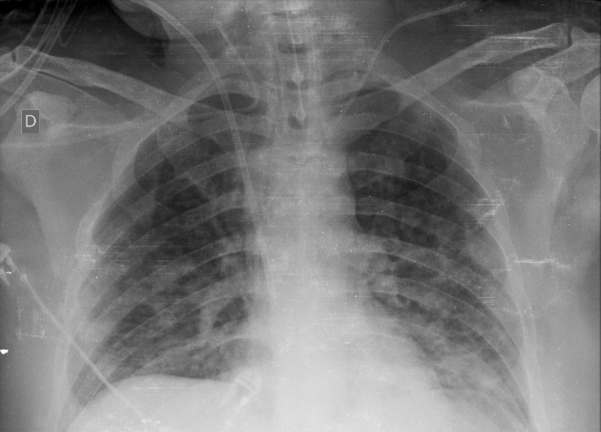
BackgroundThe COVID-19 pandemic is a global healthcare emergency. Prediction models for COVID-19 imaging are rapidly being developed to support medical decision making in imaging. However, inadequate availability of a diverse annotated dataset has limited the performance and generalizability of existing models. Purpose To create the first multi-institutional, multi-national expert annotated COVID-19 imaging dataset made freely available to the machine learning community as a research and educational resource for COVID-19 chest imaging. The Radiological Society of North America (RSNA) assembled the RSNA International COVID-19 Open Radiology Database (RICORD) collection of COVID-related imaging datasets and expert annotations to support research and education. RICORD data will be incorporated in the Medical Imaging and Data Resource Center (MIDRC), a multi-institutional research data repository funded by the National Institute of Biomedical Imaging and Bioengineering of the National Institutes of Health. Materials and Methods This dataset was created through a collaboration between the RSNA and Society of Thoracic Radiology (STR). Clinical annotation by thoracic radiology subspecialists was performed for all COVID positive chest radiography (CXR) imaging studies using a labeling schema based upon guidelines for reporting classification of COVID-19 findings in CXRs (see Review of Chest Radiograph Findings of COVID-19 Pneumonia and Suggested Reporting Language, Journal of Thoracic Imaging). Results The RSNA International COVID-19 Open Annotated Radiology Database (RICORD) consists of 998 chest x-rays from 361 patients at four international sites annotated with diagnostic labels. Patient Selection: Patients at least 18 years in age receiving positive diagnosis for COVID-19. Data Abstract
Research Benefits RICORD is available for non-commercial use (and further enrichment) by the research and education communities which may include development of educational resources for COVID-19, use of RICORD to create AI systems for diagnosis and quantification, benchmarking performance for existing solutions, exploration of distributed/federated learning, further annotation or data augmentation efforts, and evaluation of the examinations for disease entities beyond COVID-19 pneumonia. Deliberate consideration of the detailed annotation schema, demographics, and other included meta-data will be critical when generating cohorts with RICORD, particularly as more public COVID-19 imaging datasets are made available via complementary and parallel efforts. It is important to emphasize that there are limitations to the clinical “ground truth” as the SARS-CoV-2 RT-PCR tests have widely documented limitations and are subject to both false-negative and false-positive results which impact the distribution of the included imaging data, and may have led to an unknown epidemiologic distortion of patients based on the inclusion criteria. These limitations notwithstanding, RICORD has achieved the stated objectives for data complexity, heterogeneity, and high-quality expert annotations as a comprehensive COVID-19 thoracic imaging data resource. |
...
