...
| Excerpt |
|---|
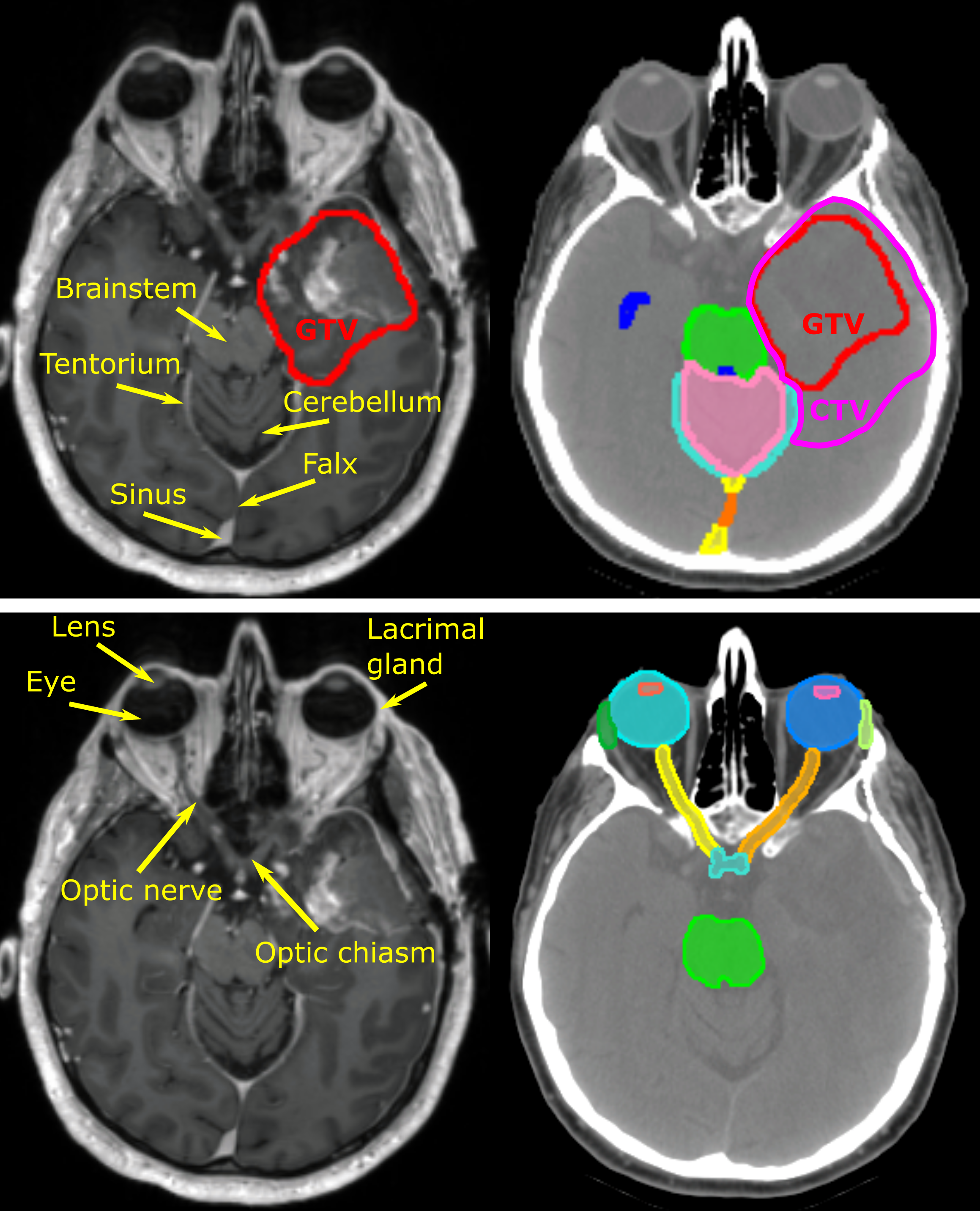
This collection consists of 230 cases of glioblastoma and low-grade glioma patients treated with surgery and adjuvant radiotherapy at Massachusetts General Hospital. The patients underwent routine post-surgical MRI examination by acquiring two MR sequences, contrast enhanced 3D-T1 and 2D multislice-T2 FLAIR required to define target volumes for radiotherapy treatment. CT scans were acquired after diagnostic imaging to use in radiotherapy treatment planning. All cases in the image set are provided with the radiotherapy targets, gross tumor volume (GTV) and clinical target volume (CTV) manually delineated by the treating radiation oncologist. The set includes glioblastoma (GBM) - 198 cases, anaplastic astrocytoma (AAC) - 23 cases, astrocytoma (AC) - 5 cases, anaplastic oligodendroglioma (AODG) - 2 cases, and oligodendroglioma (ODG) - 2 case. These abbreviations are included in the case ID. A For all cases, manual delineations are provided for the RT targets (GTV and CTV) and for organs at risk, the brainstem, optic chiasm, optic nerves, eyes, cochleae, and lacrimal glands. A subset of these 230 cases consisting of 75 cases was used for the International Challenge “Anatomical Brain Barriers to Cancer Spread: Segmentation from CT and MR Images”, ABCs, organized in conjunction with the MICCAI 2020 conference (https://abcs.mgh.harvard.edu). For these cases, manual delineations are provided including 17 structuresfor the structures used for automated definition of the CTV: the falx cerebri, tentorium cerebelli, transverse and sagittal brain sinuses, ventricles, cerebellum, brainstem, optic chiasm, optic nerves, eyes, cochlea, and lacrimal glands.. The images and manually delineated structures are to be used to develop methods for computer assisted radiotherapy target definition, algorithms for auto-delineation of the normal anatomical structures to be used for radiotherapy treatment plan optimization, and methods that utilize multi-modality images for deep learning-based image segmentation. |
...