To promote research on machine learning-based automated tumor lesion segmentation on whole-body FDG-PET/CT data we host the autoPET challenge and provide a large, publicly available training data set on Participate in this challenge via the official challenge website: https://autopet.grand-challenge.org/ |
Positron Emission Tomography / Computed Tomography (PET/CT) is an integral part of the diagnostic workup for various malignant solid tumor entities. Due to its wide applicability, Fluorodeoxyglucose (FDG) is the most widely used PET tracer in an oncological setting reflecting glucose consumption of tissues, e.g. typically increased glucose consumption of tumor lesions.
As part of the clinical routine analysis, PET/CT is mostly analyzed in a qualitative way by experienced medical imaging experts. Additional quantitative evaluation of PET information would potentially allow for more precise and individualized diagnostic decisions.
A crucial initial processing step for quantitative PET/CT analysis is segmentation of tumor lesions enabling accurate feature extraction, tumor characterization, oncologic staging and image-based therapy response assessment. Manual lesion segmentation is however associated with enormous effort and cost and is thus infeasible in clinical routine. Automation of this task is thus necessary for widespread clinical implementation of comprehensive PET image analysis.
Recent progress in automated PET/CT lesion segmentation using deep learning methods has demonstrated the principle feasibility of this task. However, despite these recent advances tumor lesion detection and segmentation in whole-body PET/CT is still a challenging task. The specific difficulty of lesion segmentation in FDG-PET lies in the fact that not only tumor lesions but also healthy organs (e.g. the brain) can have significant FDG uptake; avoiding false positive segmentations can thus be difficult. One bottleneck for progress in automated PET lesion segmentation is the limited availability of training data that would allow for algorithm development and optimization.
AutoPET is hosted at the MICCAI 2022: ![]()
and supported by the European Society for hybrid, molecular and translational imaging (ESHI)
Automatic tumor lesion segmentation in whole-body FDG-PET/CT on large-scale database of 1014 studies of 900 patients (training database) acquired on a single site:
Testing will be performed on 200 studies (held-out test database) with 100 studies originating from the same hospital as the training database and 100 are drawn from a different hospital with similar acquisition protocol to assess algorithm robustness and generalizability.
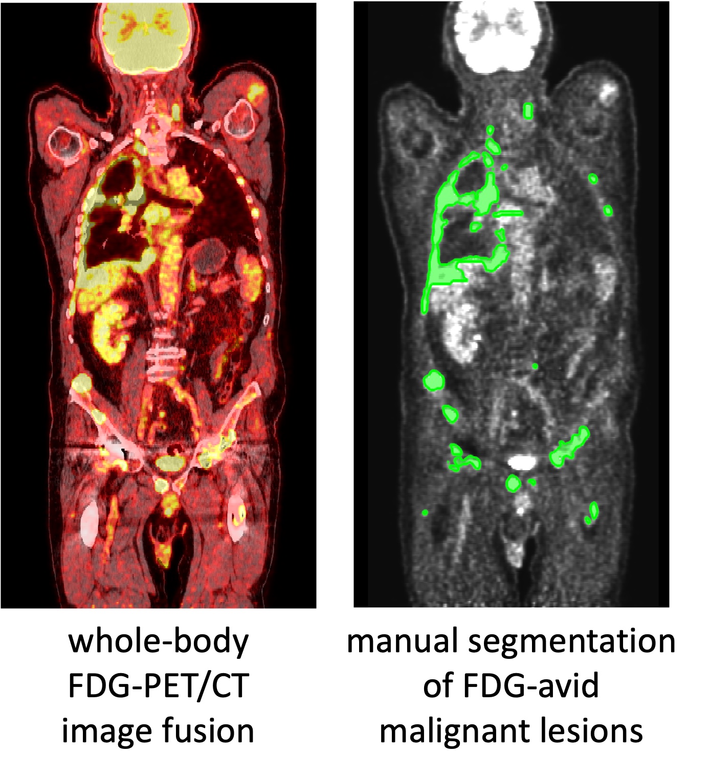
Figure: Example case of fused FDG-PET/CT whole-body data. The right image shows the manually segmented malignant lesions.