Data Access
| Data Type | Download all or Query/Filter | License |
|---|
| Images and Segmentations (DICOM, 40.7GB) |
(Download requires the NBIA Data Retriever) | | | Clinical Data (CSV, 82 kB) | | |
Click the Versions tab for more info about data releases. 
|
Detailed Description
| |
|---|
Modalities | CT, SEG | Number of Participants | 210 | Number of Studies | 210 | Number of Series | 621 | Number of Images | 71,423 | | Image Size (GB) | 40.7 |
Note: The segmentation corresponds to the arterial phase in every case. No processing or analysis was done on the other phases.
|
Citations & Data Usage Policy 
Heller, N., Sathianathen, N., Kalapara, A., Walczak, E., Moore, K., Kaluzniak, H., Rosenberg, J., Blake, P., Rengel, Z., Oestreich, M., Dean, J., Tradewell, M., Shah, A., Tejpaul, R., Edgerton, Z., Peterson, M., Raza, S., Regmi, S., Papanikolopoulos, N., Weight, C. (2019) Data from C4KC-KiTS [Data set]. The Cancer Imaging Archive. DOI: 10.7937/TCIA.2019.IX49E8NX |
Heller, N., Isensee, F., Maier-Hein, K. H., Hou, X., Xie, C., Li, F., Nan, Y., Mu, G., Lin, Z., Han, M., Yao, G., Gao, Y., Zhang, Y., Wang, Y., Hou, F., Yang, J., Xiong, G., Tian, J., Zhong, C., … Weight, C. (2021). The state of the art in kidney and kidney tumor segmentation in contrast-enhanced CT imaging: Results of the KiTS19 challenge. Medical Image Analysis, 67, 101821. https://doi.org/10.1016/j.media.2020.101821 |
Clark K, Vendt B, Smith K, Freymann J, Kirby J, Koppel P, Moore S, Phillips S, Maffitt D, Pringle M, Tarbox L, Prior F. (2013) The Cancer Imaging Archive (TCIA): Maintaining and Operating a Public Information Repository, Journal of Digital Imaging, Volume 26, Number 6, December, 2013, pp 1045-1057. DOI: 10.1007/s10278-013-9622-7 |
Other Publications Using This DataTCIA maintains a list of publications which leverage our data. If you have a publication you'd like to add please contact the TCIA Helpdesk. |
Version 3 (Current): Updated 2020/06/18| Data Type | Download all or Query/Filter |
|---|
Images and Segmentations (DICOM, 40.7GB) | | | Clinical Data (CSV, 82 kB) | |
Upon initial publication of this dataset the segmentations were stored as sagittal series, while the CT images are axial. This caused difficulties loading this dataset into various DICOM tools. Those segmentations have now been converted (in a lossless fashion) to axial to resolve these issues.
Version 2: Updated 2020/03/23
| Data Type | Download all or Query/Filter |
|---|
Images and Segmentations (DICOM, 40.7GB) | Unavailable, see version 3 note. | | Clinical Data (CSV, 82 kB) | | Added clinical data spreadsheet. Version 1: Updated 2019/12/18 | Data Type | Download all or Query/Filter |
|---|
Images (DICOM, 40.7GB) | Unavailable, see version 3 note. |
|
|
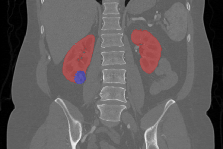
 This collection contains CT scans and segmentations from subjects from the training set of the 2019 Kidney and Kidney Tumor Segmentation Challenge (KiTS19). The challenge aimed to accelerate progress in automatic 3D semantic segmentation by releasing a dataset of CT scans for 210 patients with manual semantic segmentations of the kidneys and tumors in the corticomedullary phase.