Summary

Magnetic Resonance Imaging (MRI) quality assessment measures were generated for 

  1. T1-weighted post-contrast axial MRI sequences from 133 TCGA-GBM subjects 
  2. T1-weighted post-contrast axial MRI sequences from 46 CPTAC-GBM subjects  
  3. Both T1- and T2-weighted axial MRI sequences from 54 TCGA-CESC subjects

All MRI scans for each cohort were downloaded as DICOM files from TCIA and then processed via MRQy to compute quality measures for (a) interrogating the presence of site- or equipment-specific variations within a cohort, and (b) quantifying the impact of MRI artifacts to determine what pre-analytical corrections are needed. The MRQy output can be easily interrogated via the associated HTML5 based front-end, allowing for real-time filtering and visualization. MRQy is available for download at: http://github.com/ccipd/MRQy.  Manifest files to download the DICOM images these results were derived from are available on the Detailed Description page.

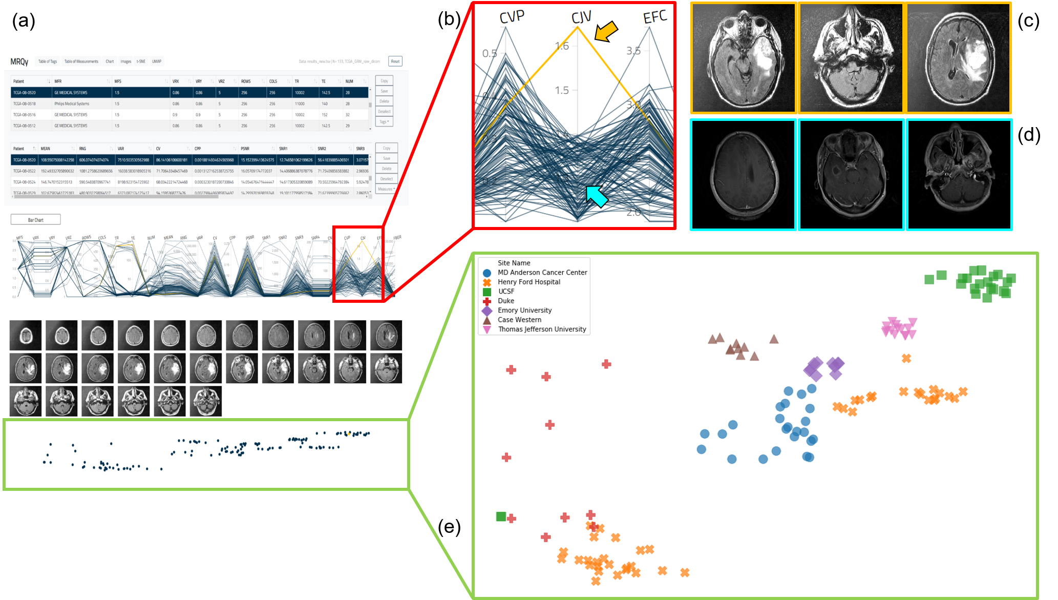
(a) MRQy front-end interface for interrogating TCGA-GBM cohort. (b) Outlier dataset identified on the parallel coordinate chart for the CJV quality measure found to exhibit shading artifacts on (c) representative images, especially when compared to (d) a different dataset without this artifact. (e) t-SNE scatter plot of quality measures revealing presence of site-specific batch effects (colors correspond to different sites, note presence of site-specific clusters).

Acknowledgements

Research reported in this publication was supported by:


Data Access


Data TypeDownload all or Query/Filter
TCGA-GBM MRQy results (XLS)

TCGA-CESC MRQy results (XLS)

CPTAC-GBM MRQy results (XLS)

Corresponding Original MR Images from TCGA-GBM (DICOM, 1.37GB)

Corresponding Original MR Images from CPTAC-GBM (DICOM, 0.4 GB)

Corresponding Original MR Images from TCGA-CESC subjects (DICOM, 2.72 GB)


Click the Versions tab for more info about data releases.

Note:   Please contact help@cancerimagingarchive.net  with any questions regarding usage.


Detailed Description

The spreadsheets provided are derived analyses from subsets of images from the TCGA-GBM, CPTAC-GBM, and TCGA-CESC collections. To download the corresponding images for your own analysis you can use the table below:


Data TypeDownload all or Query/Filter
TCGA-GBM MRQy Images (DICOM)

TCGA-CESC MRQy Images (DICOM)

CPTAC-GBM MRQy Images (DICOM)


Please be sure to cite each Collection you use according to the Citations & Data Usage policy outlined on each Collection's page.  If you have any questions or issues, please contact help@cancerimagingarchive.net.


Citations & Data Usage Policy

Sadri, A. R., Janowczyk, A., Verma, R., Antunes, J., Madabhushi, A., Tiwari, P., & Viswanath, S. E. MRQy quality measures for TCIA MRI datasets. The Cancer Imaging Archive. https://doi.org/10.7937/K9/TCIA.2020.JHZ2-T694


Clark K, Vendt B, Smith K, Freymann J, Kirby J, Koppel P, Moore S, Phillips S, Maffitt D, Pringle M, Tarbox L, Prior F. The Cancer Imaging Archive (TCIA): Maintaining and Operating a Public Information Repository, Journal of Digital Imaging, Volume 26, Number 6, December, 2013, pp 1045-1057. DOI: 10.1007/s10278-013-9622-7


Other Publications Using This Data

TCIA maintains a list of publications which leverage TCIA data. If you have a manuscript you'd like to add please contact the TCIA Helpdesk.


Version 1 (Current): Updated 2020/07/16


Data TypeDownload all or Query/Filter
TCGA-GBM MRQy results (XLS)

TCGA-CESC MRQy results (XLS)

CPTAC-GBM MRQy results (XLS)