Summary
The imaging data consists of 230 cases of glioblastoma and low-grade glioma patients treated with surgery and adjuvant radiotherapy at Massachusetts General Hospital. The patients underwent routine post-surgical MRI examination by acquiring two MR sequences, contrast enhanced 3D-T1 and 2D multislice-T2 FLAIR required to define target volumes for radiotherapy treatment. CT scans were acquired after diagnostic imaging to use in radiotherapy treatment planning. All cases in the image set are provided with the radiotherapy targets, gross tumor volume (GTV) and clinical target volume (CTV) manually delineated by the treating radiation oncologist. The subset of these 230 cases consisting of 75 cases was used for the International Challenge “Anatomical Brain Barriers to Cancer Spread: Segmentation from CT and MR Images”, ABCs, organized in conjunction with the MICCAI 2020 conference (https://abcs.mgh.harvard.edu). For these cases, manual delineations are provided including 17 structures: the falx cerebri, tentorium cerebelli, transverse and sagittal brain sinuses, ventricles, cerebellum, brainstem, optic chiasm, optic nerves, eyes, cochlea, and lacrimal glands. The set includes glioblastoma (GBM) - 198 cases, anaplastic astrocytoma (AAC) - 23 cases, astrocytoma (AC) - 5 cases, anaplastic oligodendroglioma (AODG) - 2 cases, and oligodendroglioma (ODG) - 2 case. These abbreviations are included in the case ID. The images and manually delineated structures are to be used to develop methods for computer assisted radiotherapy target definition, algorithms for auto-delineation of the normal anatomical structures to be used for radiotherapy treatment plan optimization, and methods that utilize multi-modality images for deep learning-based image segmentation.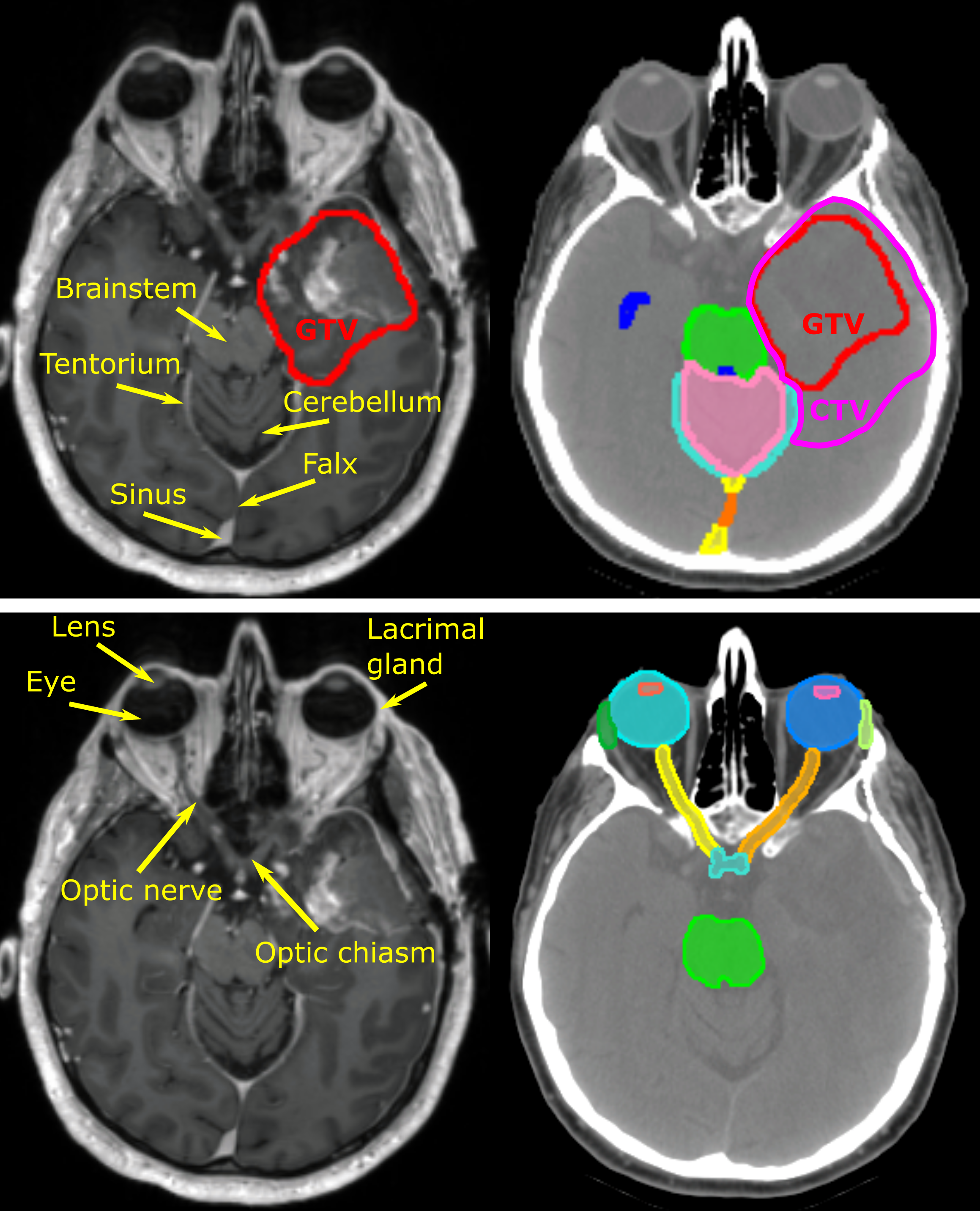
Acknowledgements
We would like to acknowledge the individuals and institutions that have provided data for this collection:
The project was supported by the Therapy Imaging Program (TIP) funded by the Federal Share of program income earned by Massachusetts General Hospital on C06 CA059267, Proton Therapy Research and Treatment Center.
Data Access
| Data Type | Download all or Query/Filter |
|---|---|
Images, Segmentations, and Radiation Therapy Structures/Doses/Plans (DICOM, XX.X GB) << latter two items only if DICOM SEG/RTSTRUCT/RTDOSE/PLAN exist >> | (Download requires the NBIA Data Retriever) |
| Tissue Slide Images (SVS, XX.X GB) | |
| Clinical data (CSV) | |
| Genomics (web) |
Click the Versions tab for more info about data releases.
Please contact help@cancerimagingarchive.net with any questions regarding usage.
Detailed Description
Image Statistics | |
|---|---|
Modalities | |
Number of Patients | |
Number of Studies | |
Number of Series | |
Number of Images | |
| Images Size (GB) |
<< Add any additional information as needed below. Likely would be something from site. >>
Citations & Data Usage Policy
Data Citation
DOI goes here. Create using Datacite with information from Collection Approval form
Publication Citation
We ask on the proposal form if they have ONE traditional publication they'd like users to cite.
Acknowledgement
Only if they ask for special acknowledgments like funding sources, grant numbers, etc in their proposal.
TCIA Citation
Clark K, Vendt B, Smith K, Freymann J, Kirby J, Koppel P, Moore S, Phillips S, Maffitt D, Pringle M, Tarbox L, Prior F. The Cancer Imaging Archive (TCIA): Maintaining and Operating a Public Information Repository, Journal of Digital Imaging, Volume 26, Number 6, December, 2013, pp 1045-1057. DOI: 10.1007/s10278-013-9622-7
Other Publications Using This Data
TCIA maintains a list of publications which leverage TCIA data. If you have a manuscript you'd like to add please contact the TCIA Helpdesk.
Version X (Current): Updated yyyy/mm/dd
| Data Type | Download all or Query/Filter |
|---|---|
| Images (DICOM, xx.x GB) | (Requires NBIA Data Retriever.) |
| Clinical Data (CSV) | Link |
| Other (format) |
<< One or two sentences about what you changed since last version. No note required for version 1. >>