...
| Excerpt |
|---|
Automated or semi-automated algorithms intended for chest CT analyses typically require the creation of a 3D map of the thoracic volume as their initial step. Identifying this anatomic region precedes fundamental tasks such as lung structure segmentation, lesion detection, and radiomics feature extraction in analysis pipelines. However, automatic approaches to segment the thoracic volume maps struggle to perform consistently in subjects with diseased lungs, yet this is exactly the circumstance for which pipeline analyses would be most useful. To address this need, we have created a dataset of thoracic volume segmentations on subjects with diseased lungs. These will help the research community compare and contrast their approaches for this foundational processing step on clinically relevant data.This dataset consists of left and right thoracic volume segmentations delineated on 402 CT scans from The Cancer Imaging Archive NSCLC Radiomics collection. Thoracic segmentations include lung parenchyma, tumor, atelectasis, and effusion when present. On scans where effusion is present, separate segmentations labeling pleural effusion alone are also provided. 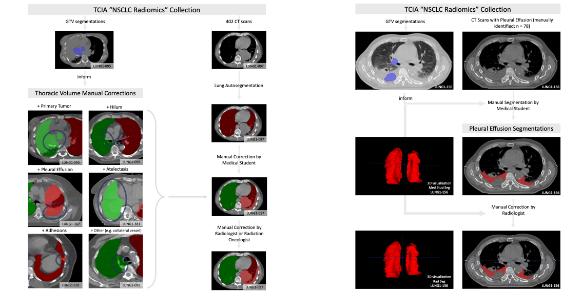 |
Acknowledgements
...