Summary


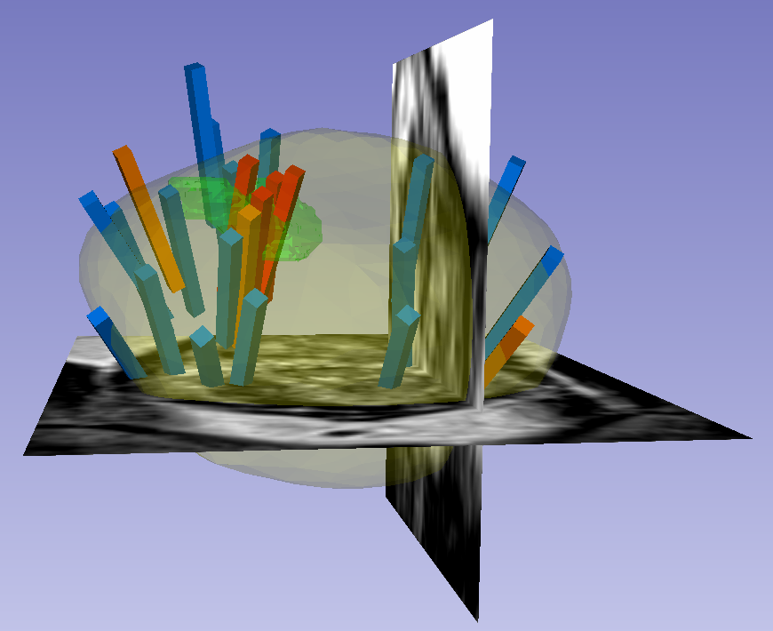
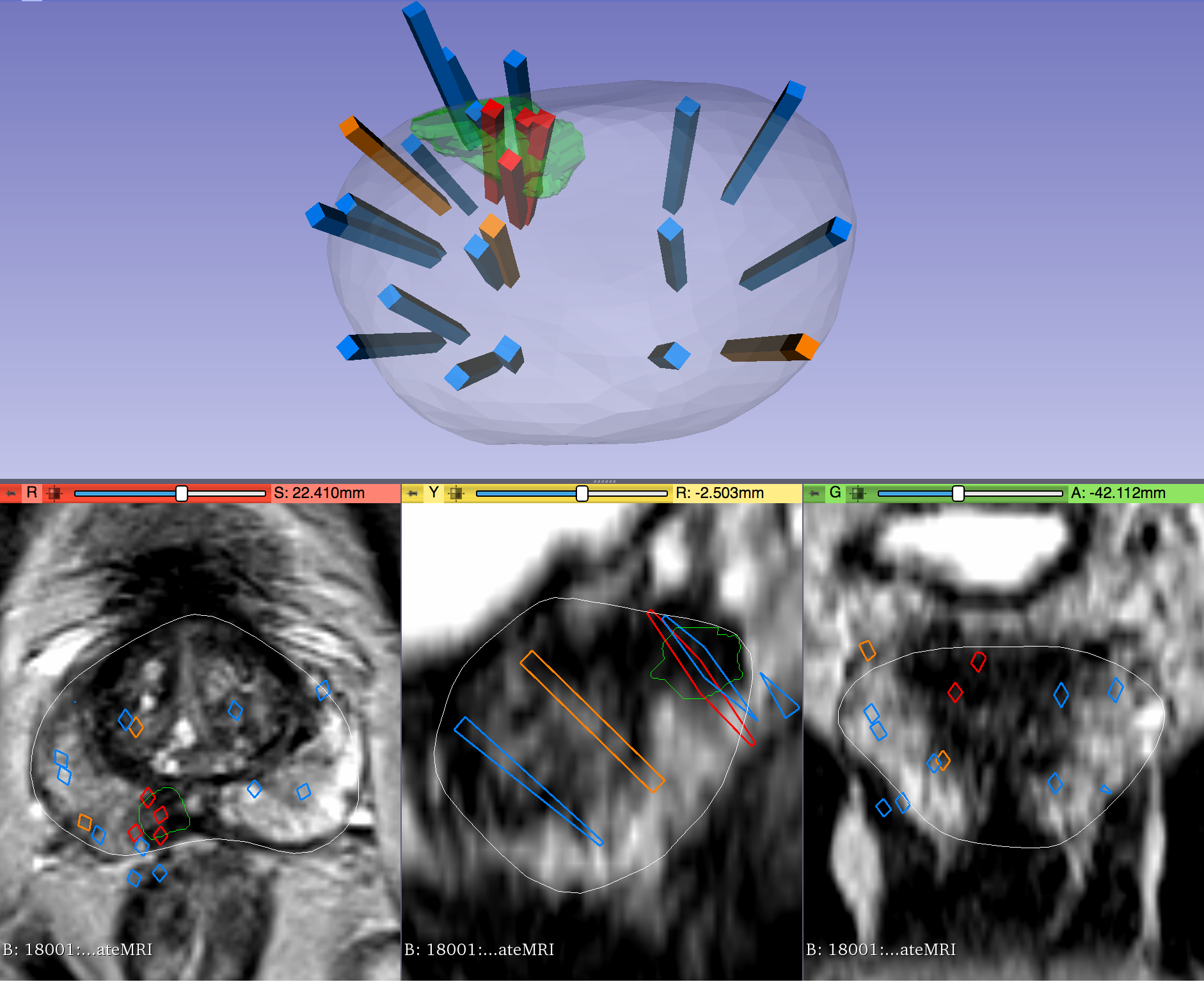
This dataset was derived from tracked biopsy sessions using the Artemis biopsy system, many of which included image fusion with MRI targets. Patients received a 3D transrectal ultrasound scan, after which nonrigid registration (e.g. “fusion”) was performed between real-time ultrasound and preoperative MRI, enabling biopsy cores to be sampled from MR regions of interest. Most cases also included sampling of systematic biopsy cores using a 12-core digital template. The Artemis system tracked targeted and systematic core locations using encoder kinematics of a mechanical arm, and recorded locations relative to the Ultrasound scan. MRI biopsy coordinates were also recorded for most cases.
...
| Localtab Group |
|---|
| Localtab |
|---|
| active | true |
|---|
| title | Data Access |
|---|
| Data AccessClick the Download button to save a ".tcia" manifest file to your computer, which you must open with the NBIA Data Retriever . Click the Search button to open our Data Portal, where you can browse the data collection and/or download a subset of its contents.
| Data Type | Download all or Query/Filter |
|---|
| Images (DICOM) 78.7 (GB) | | Target Data (XLSX) 138 (KB) | | | Biopsy Data (XLSX) 4.4 (MB) | | | |
Click the Versions tab for more info about data releases.
|
| Localtab |
|---|
| title | Detailed Description |
|---|
| Detailed Description
Image Statistics |
|
|---|
Modalities | MR, US | Number of Participants | 1151 | Number of Studies | 2795 | Number of Series | 2795 | Number of Images | 61,135 | | Images Size (GB) | 78.7 |
|
| Localtab |
|---|
| title | Citations & Data Usage Policy |
|---|
| Citations & Data Usage Policy| Public collection license |
|---|
| Info |
|---|
| Natarajan, S., Priester, A., Margolis, D., Huang, J., & Marks, L. (2020). Prostate MRI and Ultrasound With Pathology and Coordinates of Tracked Biopsy [Data set]. The Cancer Imaging Archive. DOI: 10.7937/TCIA.2020.A61IOC1A |
| Info |
|---|
| title | Publication Citation |
|---|
| Sonn, Geoffrey A., Shyam Natarajan, Daniel JA Margolis, Malu MacAiran, Patricia Lieu, Jiaoti Huang, Frederick J. Dorey, and Leonard S. Marks. "Targeted biopsy in the detection of prostate cancer using an office based magnetic resonance ultrasound fusion device." The Journal of U rology 189, no. 1 (2013): 86-92 |
| Info |
|---|
| Clark K, Vendt B, Smith K, Freymann J, Kirby J, Koppel P, Moore S, Phillips S, Maffitt D, Pringle M, Tarbox L, Prior F. The Cancer Imaging Archive (TCIA): Maintaining and Operating a Public Information Repository, Journal of Digital Imaging, Volume 26, Number 6, December, 2013, pp 1045-1057. DOI: 10.1007/s10278-013-9622-7 |
Other Publications Using This DataTCIA maintains a list of publications which leverage TCIA data. If you have a manuscript you'd like to add please contact the TCIA Helpdesk. |
| Localtab |
|---|
| Version 1 (Current): 2020/04/20
| Data Type | Download all or Query/Filter |
|---|
| Images (DICOM) 78.7 (GB) | | | Target Data (XLSX) 138 (KB) | | | Biopsy Data (XLSX) 4.4 (MB) | | | |
Added new subjects.
|
|
...

