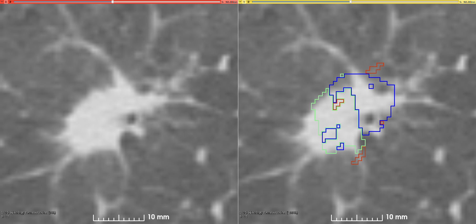
Summary This is the first attempt of mapping the extent of Invasive Adenocarcinoma onto in vivo lung CT. The mappings constitute ground truth of disease and may be used to further investigate the imaging signatures of Invasive Adenocarcinoma in ground glass pulmonary nodules. Patient with small ground glass nodules with >2 histology slices per nodule were included. Patients with solid large nodules (>40mm), with <3 histology slices or with histology slices showing substantial artifacts were excluded from this study (see reference below for details). Data collection and analysis was provided by Case Western Reserve University. This is the first attempt of mapping the extent of Invasive Adenocarcinoma onto in vivo lung CT. The mappings constitute ground truth of disease and may be used to further investigate the imaging signatures of Invasive Adenocarcinoma in ground glass pulmonary nodules. Patient with small ground glass nodules with >2 histology slices per nodule were included. Patients with solid large nodules (>40mm), with <3 histology slices or with histology slices showing substantial artifacts were excluded from this study (see reference below for details). Data collection and analysis was provided by Case Western Reserve University.
References - All the program scripts that were used for generating the results and data in this paper have been made available at https://github.com/mirabelarusu/RadPathFusionLung
- This study is described in detail in the following publication:
- Rusu M., Rajiah P., Gilkeson R., Yang M., Donatelli C., Thawani R., Jacono F.J., Linden P., Madabushi A. (2017) Co-registration of pre-operative CT with ex vivo surgically excised ground glass nodules to define spatial extent of invasive adenocarcinoma on in vivo imaging: a proof-of-concept study. European Radiology 27:10, 4209:4217. DOI: https://doi.org/10.1007/s00330-017-4813-0
| 