Child pages
  • Ultrasound data of a variety of liver masses (B-mode-and-CEUS-Liver)

Versions Compared

Key

  • This line was added.
  • This line was removed.
  • Formatting was changed.

Redirect
delay5
locationhttps://www.cancerimagingarchive.net/collection/b-mode-and-ceus-liver/
Summary

Image Added

Excerpt

Data

...

were generated as part of two ongoing clinical trials investigating the use of

...

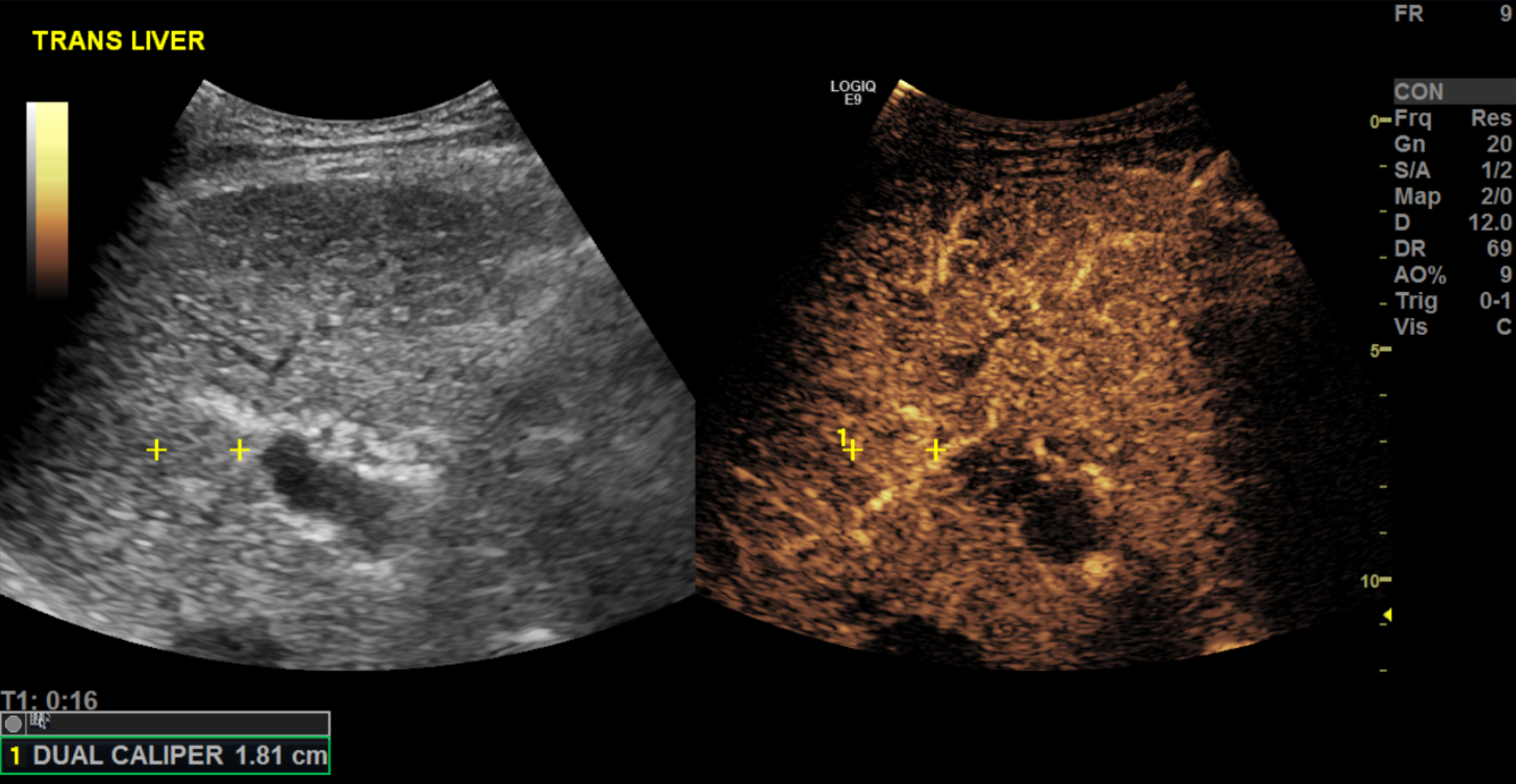
contrast-enhanced ultrasound to a) characterize indeterminate liver lesions and b) monitor treatment response to loco regional therapy. Ultrasound data was obtained on a variety of state of the art ultrasound scanners with curvilinear probes. Gain, dynamic range, focus position and depth were optimized for image quality by the performing sonographer. Images of the mass in both sagittal and transverse planes were obtained and saved in DICOM format. Full cine-loops of the contrast enhanced ultrasound are also saved in DICOM format. The reference standard used for lesion characterization included tissue pathology and contrast-enhanced cross-sectional imaging within 1 month of the ultrasound exam. The initial treatment response to transarterial chemoembolization is also available for many hepatocellular carcinoma (HCC) cases and uses pathology, retreatment angiography, or longer-term tumor response on cross-sectional imaging as a reference standard.

We expect these images can be used in a wide variety of image processing application. We are currently exploring a variety of automated intelligence algorithms for AI-based lesion characterization. Algorithms for object detection and segmentation may also be of interest. As the studies that have generated this data are also ongoing, it is expected that we can add volumetric data, contrast-enhanced ultrasound cine loops, and longer-term treatment response data to this data set in the future.


Acknowledgements

We would like to acknowledge the individuals and institutions that have provided data for this collection:

...

  • Thomas Jefferson University, Philadelphia, PA, USA - Special thanks to

...

  • John Eisenbrey,

...

  • PhD from the Department of Radiology, Andrej Lyshchik, MD,

...

  • PhD from the Department of

...

  • Radiology, Corinne Wessner, MBA from the Department of Radiology

  • This work was supported in whole or in part under R01 CA194307 ; R01 CA215520 .
Localtab Group


Localtab
activetrue
titleData Access

Data Access

...

Data

...

TypeDownload all or Query/FilterLicense
Images (DICOM,

...

Image Removed  Image Removed

...

Image Removed

Click the Versions tab for more info about data releases.

253 GB)


Tcia button generator
urlhttps://wiki.cancerimagingarchive.net/download/attachments/61080617/B-mode-and-CEUS-Liver_20220218.tcia?api=v2


Tcia button generator
labelSearch
urlhttps://www.cancerimagingarchive.net/nbia-search/?CollectionCriteria=B-mode-and-CEUS-Liver


(Download requires NBIA Data Retriever)

Tcia cc by 4

Clinical data (XLSX, 24 kB)


Tcia button generator
urlhttps://wiki.cancerimagingarchive.net/download/attachments/61080617/B-mode-and-CEUS-Liver_ReferenceStandards_v2_20220218.xlsx?api=v2


Tcia cc by 4


Nci_crdc additional resources


Localtab
titleDetailed Description

Detailed Description

Image Statistics


Modalities

US

...

Number of Participants

...

120

Number of Studies

...

120

Number of Series

120

Number of Images

1859

Images Size (GB)

...

253


Localtab
titleCitations & Data Usage Policy

Citations & Data Usage Policy

Add any special restrictions in here.

...

titleAcknowledgement

...

tcia-limited-license-policy

Info
titleData Citation

...

DOI goes here. Create using pubhub with information from Collection Approval form

Eisenbrey, J., Lyshchik, A., & Wessner, C. (2021). Ultrasound data of a variety of liver masses [Data set]. The Cancer Imaging Archive.  DOI: https://doi.org/10.7937/TCIA.2021.v4z7-tc39


Info
titleTCIA Citation

Clark K, Vendt B, Smith K, Freymann J, Kirby J, Koppel P, Moore S, Phillips S, Maffitt D, Pringle M, Tarbox L, Prior F. The Cancer Imaging Archive (TCIA): Maintaining and Operating a Public Information Repository, Journal of Digital Imaging, Volume 26, Number 6, December, 2013, pp 1045-1057. DOI: 10.1007/s10278-013-9622-7

Other Publications Using This Data

TCIA maintains a list of publications which leverage TCIA data. If you have a manuscript you'd like to add please contact the TCIA Helpdesk.


Localtab
titleVersions

Version

...

2 (Current): Updated

...

2022/

...

02/

...

18

Data TypeDownload all or Query/Filter
Images (DICOM,

...

Image Removed Image Removed

(Requires NBIA Data Retriever .)

...

Image Removed

253 GB)


Tcia button generator
urlhttps://wiki.cancerimagingarchive.net/download/attachments/61080617/B-mode-and-CEUS-Liver_20220218.tcia?api=v2


Tcia button generator
labelSearch
urlhttps://www.cancerimagingarchive.net/nbia-search/?CollectionCriteria=B-mode-and-CEUS-Liver


(Download requires NBIA Data Retriever app)

Clinical data (XLSX, 24KB)


Tcia button generator
urlhttps://wiki.cancerimagingarchive.net/download/attachments/61080617/B-mode-and-CEUS-Liver_ReferenceStandards_v2_20220218.xlsx?api=v2


Added 45 new participants and a new spreadsheet version.

Version 1: Updated 2021/08/04

Data TypeDownload all or Query/Filter
Images (DICOM, 208 GB)


Tcia button generator
urlhttps://wiki.cancerimagingarchive.net/download/attachments/61080617/B-mode-and-CEUS-Liver_20210728.tcia?api=v2


(Download requires NBIA Data Retriever app)

Clinical data (XLSX, 19KB)


Tcia button generator
urlhttps://wiki.cancerimagingarchive.net/download/attachments/61080617/B-mode-and-CEUS-Liver_ReferenceStandards_v1_20210804.xlsx?api=v2


7/28/21 Added new participants.
8/4/21 Added spreadsheet

Added new subjects.

Version 1: Updated 2018/10/24

...

.