Summary
Redirect delay 5 location https://www.cancerimagingarchive.net/collection/ucsf-pdgm/
Introduction
| delay | 5 |
|---|---|
| location | https://www.cancerimagingarchive.net/collection/ucsf-pdgm/ |
| Column | ||
|---|---|---|
| ||
|
MRI-based artificial intelligence (AI) research on patients with brain gliomas has been rapidly increasing in popularity in recent years in part due to a growing number of publicly available MRI datasets. Notable examples include The Cancer Genome Atlas Glioblastoma dataset (TCGA-GBM) consisting of 262 subjects and the International Brain Tumor Segmentation (BraTS) challenge dataset consisting of 542 subjects (including 243 preoperative cases from TCGA-GBM). The public availability of these glioma MRI datasets has fostered the growth of numerous emerging AI techniques including automated tumor segmentation, radiogenomics, and MRI-based survival prediction. Despite these advances, existing publicly available glioma MRI datasets have been largely limited to only 4 MRI contrasts (T2, T2/FLAIR, and T1 pre- and post-contrast) and imaging protocols vary significantly in terms of magnetic field strength and acquisition parameters. Here we present the University of California San Francisco Preoperative Diffuse Glioma MRI (UCSF-PDGM) dataset. The UCSF-PDGM dataset includes 501 subjects with histopathologically-proven diffuse gliomas who were imaged with a standardized 3 Tesla preoperative brain tumor MRI protocol featuring predominantly 3D imaging, as well as advanced diffusion and perfusion imaging techniques. The dataset also includes isocitrate dehydrogenase (IDH) mutation status for all cases and O[6]-methylguanine-DNA methyltransferase (MGMT) promotor methylation status for World Health Organization (WHO) grade III and IV gliomas. The UCSF-PDGM has been made publicly available in the hopes that researchers around the world will use these data to continue to push the boundaries of AI applications for diffuse gliomas.
Methods
Patient Population
Data collection was performed in accordance with relevant guidelines and regulations and was approved by the University of California San Francisco institutional review board with a waiver for consent. The dataset population consisted of 501* adult patients with histopathologically confirmed grade II-IV diffuse gliomas who underwent preoperative MRI, initial tumor resection, and tumor genetic testing at a single medical center between 2015 and 2021. Patients with any prior history of brain tumor treatment were excluded; however, history of tumor biopsy was not considered an exclusion criterion.
Genetic Biomarker Testing
All subjects’ tumors were tested for IDH mutations by genetic sequencing of tissue acquired during biopsy or resection. All grade III and IV tumors were tested for MGMT methylation status using a methylation sensitive quantitative PCR assay.
Study participant demographic data
The 501* cases included in the UCSF-PDGM include 55 (11%) grade II, 42 (9%) grade III, and 403 (80%) grade IV tumors. There was a male predominance for all tumor grades (56%, 60%, and 60%, respectively for grades II-IV). IDH mutations were identified in a majority of grade II (83%) and grade III (67%) tumors and a small minority of grade IV tumors (8%). MGMT promoter hypermethylation was detected in 63% of grade IV gliomas and was not tested for in a majority of lower grade gliomas. 1p/19q codeletion was detected in 20% of grade II tumors and a small minority of grade III (5%) and IV (<1%) tumors. Tabulated details and glossary are available in the Data Access and Detailed Description tabs below.
Image Acquisition
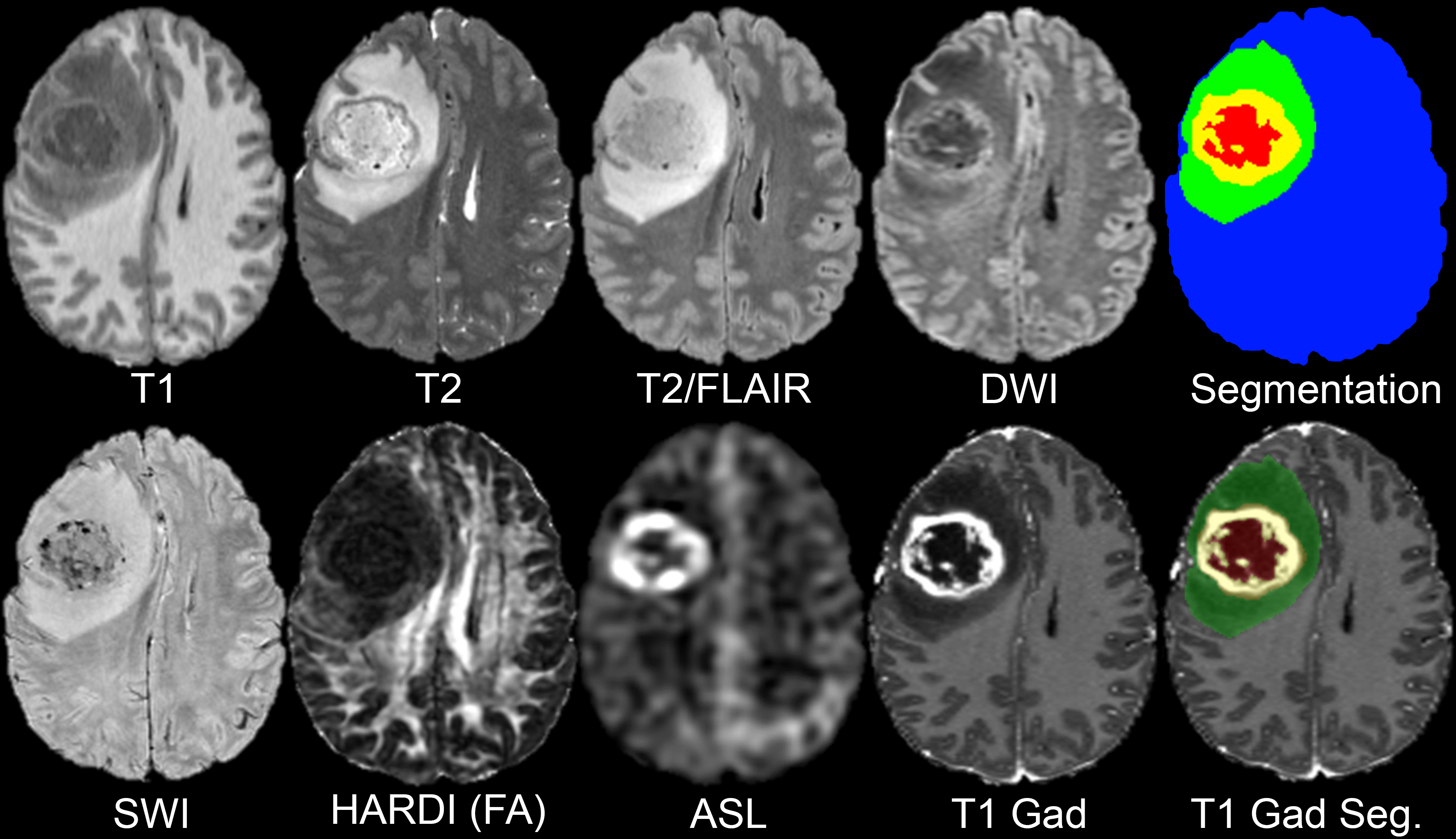
All preoperative MRI was performed on a 3.0 tesla scanner (Discovery 750, GE Healthcare, Waukesha, Wisconsin, USA) and a dedicated 8-channel head coil (Invivo, Gainesville, Florida, USA). The imaging protocol included 3D T2-weighted, T2/FLAIR-weighted, susceptibility-weighted (SWI), diffusion-weighted (DWI), pre- and post-contrast T1-weighted images, 3D arterial spin labeling (ASL) perfusion images, and 2D 55-direction high angular resolution diffusion imaging (HARDI). Over the study period, two gadolinium-based contrast agents were used: gadobutrol (Gadovist, Bayer, LOC) at a dose of 0.1 mL/kg and gadoterate (Dotarem, Guerbet, Aulnay-sous-Bois, France) at a dose of 0.2 mL/kg.
Image Pre-Processing
HARDI data were eddy current corrected and processed using the Eddy and DTIFIT modules from FSL 6.0.2 yielding isotropic diffusion weighted images (DWI) and several quantitative diffusivity maps: mean diffusivity (MD), axial diffusivity (AD), radial diffusivity (RD), and fractional anisotropy (FA). Eddy correction was performed with outlier replacement on and topup correction off. DTIFIT was performed with simple least squares regression. Each image contrast was registered and resampled to the 3D space defined by the T2/FLAIR image (1 mm isotropic resolution) using automated non-linear registration (Advanced Normalization Tools). Resampled co-registered data were then skull stripped using a previously described and publicly available deep-learning algorithm: https://www.github.com/ecalabr/brain_mask/.
Tumor Segmentation
Multicompartment tumor segmentation of study data was undertaken as part of the 2021 BraTS challenge. Briefly, image data first underwent automated segmentation using an ensemble model consisting of prior BraTS challenge winning segmentation algorithms. Images were then manually corrected by trained radiologists and approved by 2 expert reviewers. Segmentation included three major tumor compartments: enhancing tumor, non-enhancing/necrotic tumor, and surrounding FLAIR abnormality (sometimes referred to as edema).
The UCSF-PDGM adds to on an existing body of publicly available diffuse glioma MRI datasets that are commonly used in AI research applications. As MRI-based AI research applications continue to grow, new data are needed to foster development of new techniques and increase the generalizability of existing algorithms. The UCSF-PDGM not only significantly increases the total number of publicly available diffuse glioma MRI cases, but also provides a unique contribution in terms of MRI technique. The inclusion of 3D sequences and advanced MRI techniques like ASL and HARDI provides a new opportunity for researchers to explore the potential utility of cutting-edge clinical diagnostics for AI applications. In addition, these advanced imaging techniques may prove useful for radiogenomic studies focused on identification of IDH mutations or MGMT promoter methylation.
The UCSF-PDGM dataset, particularly when combined with existing publicly available datasets, has the potential to fuel the next phase of radiologic AI research on diffuse gliomas. However, the UCSF-PDGM dataset’s potential will only be realized if the radiology AI research community takes advantage of this new data resource. We hope that this dataset sparks inspiration in the next generation of AI researchers, and we look forward to the new techniques and discoveries that the UCSF-PDGM will generate.
Acknowledgements
We would like to acknowledge the individuals and institutions that have provided data for this collection:
Research was supported by the National Institutes of Health Ruth L. Kirschstein Institutional National Research Service Award under award number T32EB001631 and by the RSNA Research & Education Foundation under grant number RR2011. The content is solely the responsibility of the authors and does not necessarily represent the official views of the RSNA R&E Foundation.
| Localtab Group | |||||||||||||||||||||||||||||||||||||||||||||||||||||||||||||||||||||||||||||||||||||||||||||||||||||||||||||||||||||||||||||||||||||||||||||||||||||||||||||||||||||||||||||||||||||||||||||||||||||||||||||||||||||||||||||||||||||||||||||||||||||
|---|---|---|---|---|---|---|---|---|---|---|---|---|---|---|---|---|---|---|---|---|---|---|---|---|---|---|---|---|---|---|---|---|---|---|---|---|---|---|---|---|---|---|---|---|---|---|---|---|---|---|---|---|---|---|---|---|---|---|---|---|---|---|---|---|---|---|---|---|---|---|---|---|---|---|---|---|---|---|---|---|---|---|---|---|---|---|---|---|---|---|---|---|---|---|---|---|---|---|---|---|---|---|---|---|---|---|---|---|---|---|---|---|---|---|---|---|---|---|---|---|---|---|---|---|---|---|---|---|---|---|---|---|---|---|---|---|---|---|---|---|---|---|---|---|---|---|---|---|---|---|---|---|---|---|---|---|---|---|---|---|---|---|---|---|---|---|---|---|---|---|---|---|---|---|---|---|---|---|---|---|---|---|---|---|---|---|---|---|---|---|---|---|---|---|---|---|---|---|---|---|---|---|---|---|---|---|---|---|---|---|---|---|---|---|---|---|---|---|---|---|---|---|---|---|---|---|---|---|---|---|---|---|---|---|---|---|---|---|---|---|---|---|---|---|---|
|