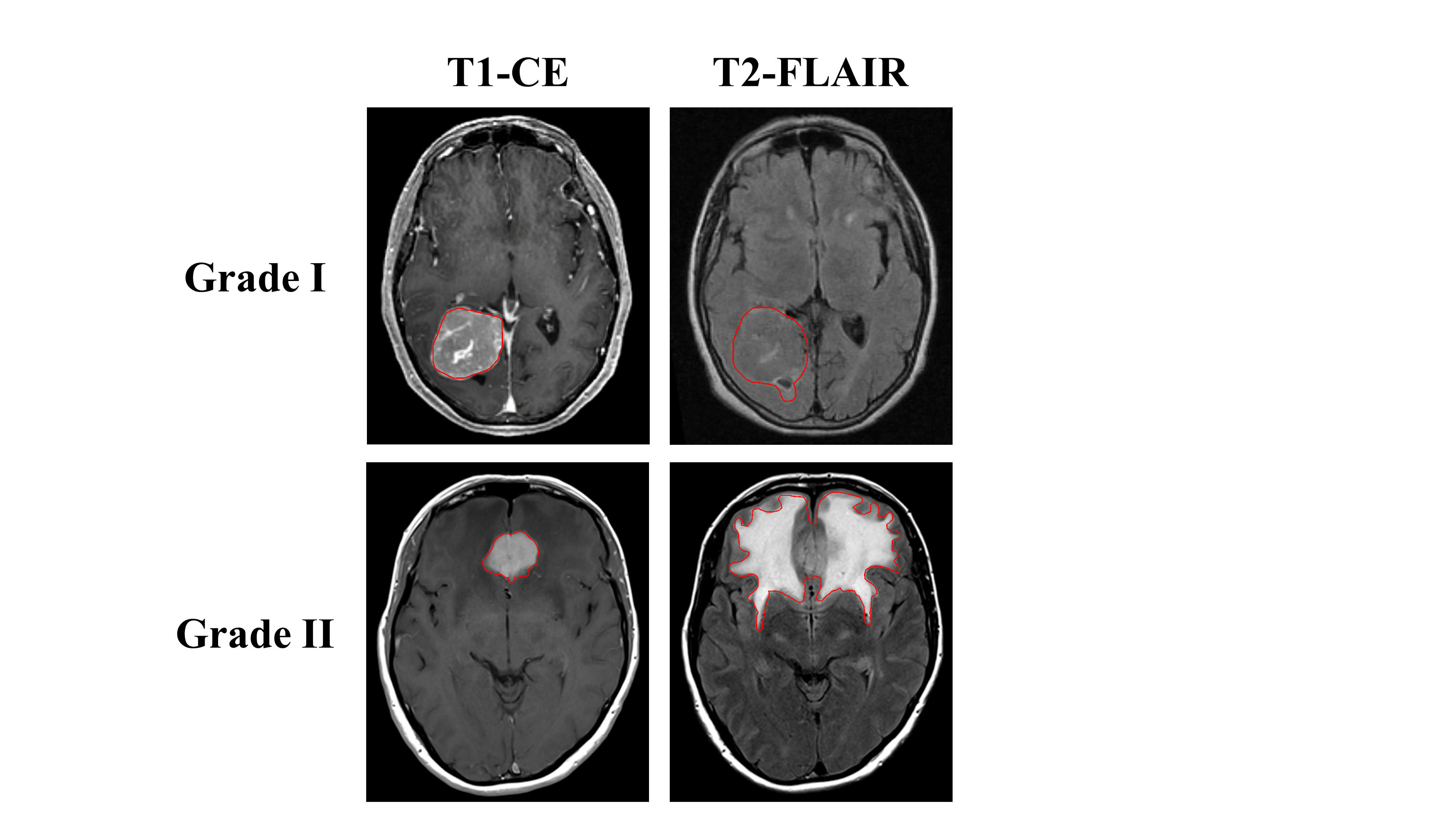
 The study included 96 consecutive treatment naïve patients with intracranial meningiomas treated with surgical resection from 2010 to 2019. All patients had pre-operative T1, T1-CE, and T2-FLAIR MR images with subsequent subtotal or gross total resection of pathologically confirmed grade I or grade II meningiomas. A neuropathology team reviewed histopathology, including two subspecialty trained neuropathologists and one neuropathology fellow. The meningioma grade was confirmed based on current classification guidelines, most recently described in the 2016 WHO Bluebook. Clinical information includes grade, subtype, type of surgery, tumor location, and atypical features. Meningioma labels on T1-CE and T2-FLAIR images will also be provided in DICOM format. The hyperintense T1-contrast enhancing tumor and hyperintense T2-FLAIR and tumor were manually contoured on each MRI and reviewed by a central nervous system radiation oncologist specialist. The study included 96 consecutive treatment naïve patients with intracranial meningiomas treated with surgical resection from 2010 to 2019. All patients had pre-operative T1, T1-CE, and T2-FLAIR MR images with subsequent subtotal or gross total resection of pathologically confirmed grade I or grade II meningiomas. A neuropathology team reviewed histopathology, including two subspecialty trained neuropathologists and one neuropathology fellow. The meningioma grade was confirmed based on current classification guidelines, most recently described in the 2016 WHO Bluebook. Clinical information includes grade, subtype, type of surgery, tumor location, and atypical features. Meningioma labels on T1-CE and T2-FLAIR images will also be provided in DICOM format. The hyperintense T1-contrast enhancing tumor and hyperintense T2-FLAIR and tumor were manually contoured on each MRI and reviewed by a central nervous system radiation oncologist specialist.
|