...
For convenience you can obtain the publications specifically based on TCIA in Endnote XML format: Pubs_basedon_TCIA_TCIA06150715.xml. This should be usable as input to your favorite reference management system.
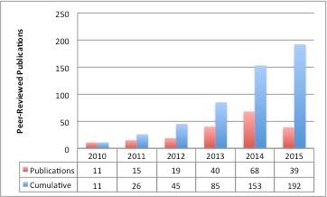
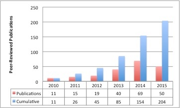
TCIA-Related Publication History
...
| Table of Contents | ||||
|---|---|---|---|---|
|
TCIA General
- Toga AW, Dinov ID. Sharing big biomedical data. Journal of Big Data. 2015;2(1):1-12.
- Herskovits EH. Quantitative Radiology: Applications to Oncology. Emerging Applications of Molecular Imaging to Oncology. 2014;124:1-30.
- Armato SG, Hadjiiski L, Tourassi GD, Drukker K, Giger ML, Li F, Redmond G, Farahani K, Kirby JS, Clarke LP. Special Section Guest Editorial: LUNGx Challenge for computerized lung nodule classification: reflections and lessons learned. Journal of Medical Imaging. 2015;2(2):020103-.
- Moore SM, Maffitt DR, Smith KE, Kirby JS, Clark KW, Freymann JB, Vendt BA, Tarbox LR, Prior FW. De-identification of Medical Images with Retention of Scientific Research Value. RadioGraphics. 2015;35(3):727-35. doi: doi:10.1148/rg.2015140244.
- Bourne PE. DOIs for DICOM Raw Images: Enabling Science Reproducibility. Radiology. 2015;275(1):3-4. doi: doi:10.1148/radiol.15150144. PubMed PMID: 25799330.
- Commean PK, Rathmell JM, Clark KW, Maffitt DR, Prior FW. A Query Tool for Investigator Access to the Data and Images of the National Lung Screening Trial. Journal of Digital Imaging. 2015:1-9
- Moore SM, Maffitt DR, Smith KE, Kirby JS, Clark KW, Freymann JB, Vendt BA, Tarbox LR, Prior FW. De-identification of Medical Images with Retention of Scientific Research Value. RadioGraphics. 2015;35(3):727-35. doi: doi:10.1148/rg.2015140244.
- Bourne PE. DOIs for DICOM Raw Images: Enabling Science Reproducibility. Radiology. 2015;275(1):3-4. doi: doi:10.1148/radiol.15150144. PubMed PMID: 25799330.
- Commean PK, Rathmell JM, Clark KW, Maffitt DR, Prior FW. A Query Tool for Investigator Access to the Data and Images of the National Lung Screening Trial. Journal of Digital Imaging. 2015:1-9. (paper)
- Gutman DA, Dunn Jr WD, Cobb J, Stoner RM, Kalpathy-Cramer J, Erickson B. Web based tools for visualizing imaging data and development of XNATView, a zero footprint image viewer. Frontiers in Neuroinformatics. 2014;8.(paper)
- Erickson BJ, Fajnwaks P, Langer SG, and Perry J. Multisite Image Data Collection and Management Using the RSNA Image Sharing Network., Translational oncology, 2014. 7(1):36-39. (paper)
- Gutman DA, Dunn Jr WD, Cobb J, Somanna D, et al. Cancer Digital Slide Archive: an informatics resource to support integrated in silico analysis of TCGA pathology data., Journal of the American Medical Informatics Association, 2013. 20(6): p. 1091-1098. doi: 10.1136/amiajnl-2012-001469 (paper)
- Clark K, Vendt B, Smith K, Freymann J, Kirby J, Koppel P, Moore S, Phillips S, Maffitt D, Pringle M, Tarbox L, Prior F. The Cancer Imaging Archive (TCIA): Maintaining and Operating a Public Information Repository, Journal of Digital Imaging, Volume 26, Number 6, December, 2013, pp 1045-1057. (paper)
- Prior FW, Clark K, Commean P, Freymann J, Jaffe C, Kirby J, Moore S, Smith K, Tarbox L, Vendt B. TCIA: an information resource to enable open science. Engineering in Medicine and Biology Society (EMBC), 2013 35th Annual International Conference of the IEEE; 2013. (paper)
- Jaffe, C Carl. Imaging and Genomics: Is There a Synergy?. Radiology. 2012. 264(2):329-31.(paper).
- Mongkolwat P, Channin DS, Kleper V, Rubin DL. Informatics in Radiology: An Open-Source and Open-Access Cancer Biomedical Informatics Grid Annotation and Image Markup Template Builder.Radiographics .2012. 32(4):1223-32. (paper).
- Freymann JB, Kirby JS, Perry JH, Clunie DA, and Jaffe CC. Image data sharing for biomedical research—meeting HIPAA requirements for de-identification.Journal of Digital Imaging 25, no. 1 (2012): 14-24. (paper)
- Villani L and Prati RC. Classificação Multirrótulo na Anotação Automática de Nódulo Pulmonar Solitário. Congresso Brasileiro de Informática em Saúde (CBIS’2012). Citado na. 2012.(paper)
Radiogenomics
Tomczak K, Czerwińska P, Wiznerowicz M. The Cancer Genome Atlas (TCGA): an immeasurable source of knowledge. Contemp Oncol (Pozn). 2015;19(1A):A68-A77.
Pope WB. Genomics of Brain Tumor Imaging. Neuroimaging Clinics of North America. 2015;25(1):105-19.
- Colen R, Foster I, Gatenby R, Giger ME, Gillies R, Gutman D, Heller M, Jain R, Madabhushi A, Madhavan S, Napel S, Rao A, Saltz J, Tatum J, Verhaak R, Whitman G. NCI Workshop Report: Clinical and Computational Requirements for Correlating Imaging Phenotypes with Genomics Signatures. Translational Oncology. 2014;7(5):556-69. doi: http://dx.doi.org/10.1016/j.tranon.2014.07.007.
- Rao A. Exploring relationships between multivariate radiological phenotypes and genetic features: A case-study in Glioblastoma using the Cancer Genome Atlas, Global Conference on Signal and Information Processing (GlobalSIP), 2013 IEEE.
- Gevaert O, Xu J, Hoang CD, Leung AN, Xu Y, Quon A, Rubin DL, Napel S, Plevritis SK. Non-small cell lung cancer: identifying prognostic imaging biomarkers by leveraging public gene expression microarray data--methods and preliminary results. Radiology. 2012;264(2):387-96. Epub 2012/06/23. doi: 10.1148/radiol.12111607. PubMed PMID: 22723499; PubMed Central PMCID: PMCPMC3401348.
Algorithm Development
- Stoner RM, Kalpathy-Cramer J, Erickson B. Web based tools for visualizing imaging data and development of XNATView, a zero footprint image viewer. Frontiers in Neuroinformatics. 2014;8.(paper)
- Erickson BJ, Fajnwaks P, Langer SG, and Perry J. Multisite Image Data Collection and Management Using the RSNA Image Sharing Network., Translational oncology, 2014. 7(1):36-39. (paper)
- Gutman DA, Cobb J, Somanna D, et al. Cancer Digital Slide Archive: an informatics resource to support integrated in silico analysis of TCGA pathology data., Journal of the American Medical Informatics Association, 2013. 20(6): p. 1091-1098. doi: 10.1136/amiajnl-2012-001469 (paper)
- Clark K, Vendt B, Smith K, Freymann J, Kirby J, Koppel P, Moore S, Phillips S, Maffitt D, Pringle M, Tarbox L, Prior F. The Cancer Imaging Archive (TCIA): Maintaining and Operating a Public Information Repository, Journal of Digital Imaging, Volume 26, Number 6, December, 2013, pp 1045-1057. (paper)
- Prior FW, Clark K, Commean P, Freymann J, Jaffe C, Kirby J, Moore S, Smith K, Tarbox L, Vendt B. TCIA: an information resource to enable open science. Engineering in Medicine and Biology Society (EMBC), 2013 35th Annual International Conference of the IEEE; 2013. (paper)
- Jaffe, C Carl. Imaging and Genomics: Is There a Synergy?. Radiology. 2012. 264(2):329-31.(paper).
- Mongkolwat P, Channin DS, Kleper V, Rubin DL. Informatics in Radiology: An Open-Source and Open-Access Cancer Biomedical Informatics Grid Annotation and Image Markup Template Builder.Radiographics .2012. 32(4):1223-32. (paper).
- Freymann JB, Kirby JS, Perry JH, Clunie DA, and Jaffe CC. Image data sharing for biomedical research—meeting HIPAA requirements for de-identification.Journal of Digital Imaging 25, no. 1 (2012): 14-24. (paper)
- Villani L and Prati RC. Classificação Multirrótulo na Anotação Automática de Nódulo Pulmonar Solitário. Congresso Brasileiro de Informática em Saúde (CBIS’2012). Citado na. 2012.(paper)
Radiogenomics
Tomczak K, Czerwińska P, Wiznerowicz M. The Cancer Genome Atlas (TCGA): an immeasurable source of knowledge. Contemp Oncol (Pozn). 2015;19(1A):A68-A77.
Pope WB. Genomics of Brain Tumor Imaging. Neuroimaging Clinics of North America. 2015;25(1):105-19.
- Colen R, Foster I, Gatenby R, Giger ME, Gillies R, Gutman D, Heller M, Jain R, Madabhushi A, Madhavan S, Napel S, Rao A, Saltz J, Tatum J, Verhaak R, Whitman G. NCI Workshop Report: Clinical and Computational Requirements for Correlating Imaging Phenotypes with Genomics Signatures. Translational Oncology. 2014;7(5):556-69. doi: http://dx.doi.org/10.1016/j.tranon.2014.07.007.
- Rao A. Exploring relationships between multivariate radiological phenotypes and genetic features: A case-study in Glioblastoma using the Cancer Genome Atlas, Global Conference on Signal and Information Processing (GlobalSIP), 2013 IEEE.
- Gevaert O, Xu J, Hoang CD, Leung AN, Xu Y, Quon A, Rubin DL, Napel S, Plevritis SK. Non-small cell lung cancer: identifying prognostic imaging biomarkers by leveraging public gene expression microarray data--methods and preliminary results. Radiology. 2012;264(2):387-96. Epub 2012/06/23. doi: 10.1148/radiol.12111607. PubMed PMID: 22723499; PubMed Central PMCID: PMCPMC3401348.
Algorithm Development
- Vallières M, Freeman C, Skamene S, El Naqa I. A radiomics model from joint FDG-PET and MRI texture features for the prediction of lung metastases in soft-tissue sarcomas of the extremities. Physics in medicine and biology. 2015;60(14):5471.
- Kazdal S, Dogan B, Camurcu AY, editors. Computer-aided detection of brain tumors using image processing techniques. Signal Processing and Communications Applications Conference (SIU), 2015 23th; 2015: IEEE.
- Gupta A, Martens O, Le Moullec Y, Saar T, editors. A tool for lung nodules analysis based on segmentation and morphological operation. Intelligent Signal Processing (WISP), 2015 IEEE 9th International Symposium on; 2015: IEEE.
- Benninghoff H, Garcke H. Segmentation of Three-dimensional Images with Parametric Active Surfaces and Topology Changes. arXiv preprint arXiv:150607136. 2015.
- Zabala-Travers S, Choi M, Cheng W-C, Badano A. Effect of color visualization and display hardware on the visual assessment of pseudocolor medical images. Medical Physics. 2015;42(6):2942-54.
- Guvenis A, Koc A. OPTIMISING DELINEATION ACCURACY OF TUMOURS IN PET FOR RADIOTHERAPY PLANNING USING BLIND DECONVOLUTION. Radiation Protection Dosimetry. 2015:ncv110.
- Grove O, Berglund AE, Schabath MB, Aerts HJ, Dekker A, Wang H, Velazquez ER, Lambin P, Gu Y, Balagurunathan Y. Quantitative Computed Tomographic Descriptors Associate Tumor Shape Complexity and Intratumor Heterogeneity with Prognosis in Lung Adenocarcinoma. PloS one. 2015;10(3).
- Buerger C, Sénégas J, Kabus S, Carolus H, Schulz H, Agarwal H, Turkbey B, Choyke P, Renisch S. Comparing nonrigid registration techniques for motion corrected MR prostate diffusion imaging. Medical physics. 2015;42(1):69-80.
- Abedini M, Codella N, Connell J, Garnavi R, Merler M, Pankanti S, Smith J, Syeda-Mahmood T. A generalized framework for medical image classification and recognition. IBM Journal of Research and Development. 2015;59(2/3):1: -: 18.
- Blessy SPS, Sulochana CH. Performance analysis of unsupervised optimal fuzzy clustering algorithm for MRI brain tumor segmentation. Technology and Health Care. 2014.
- ElNawasany AM, Ali AF, Waheed ME. A Novel Hybrid Perceptron Neural Network Algorithm for Classifying Breast MRI Tumors. Advanced Machine Learning Technologies and Applications: Springer; 2014. p. 357-66.
- Hong S, Huang Y, Cao Y, Chen X, Han J-DJ. Approaches to uncovering cancer diagnostic and prognostic molecular signatures. Molecular & Cellular Oncology. 2014.
- Codella N, Connell J, Pankanti S, Merler M, and Smith JR. Automated Medical Image Modality Recognition by Fusion of Visual and Text Information. Medical Image Computing and Computer-Assisted Intervention. 2014, Springer. 487-495. (link)
- Ertugrul OF. Adaptive Texture Energy Measure Method. International Journal of Intelligent Information Systems. 2014. 3(2):13-18. doi:10.11648/j.ijiis.20140302.11 (link)
- Kawa J, Juszczyk J, Pyciński B, Badura P, Pietka E. Radiological Atlas for Patient Specific Model Generation. Information Technologies in Biomedicine, 2014 4:69-82. 10.1007/978-3-319-06596-0_7. (link)
- Kowalik-Urbaniak I, Brunet D, Wang J, Koff D, Smolarski-Koff N, Vrscay ER, Wallace B, Wang Z.The quest for ‘diagnostically lossless’ medical image compression: a comparative study of objective quality metrics for compressed medical images. SPIE Medical Imaging. 2014. Vol. 9073. International Society for Optics and Photonics. doi:10.1117/12.2043196 (link)
- Naresh P and Shettar R. Image Processing and Classification Techniques for Early Detection of Lung Cancer for Preventive Health Care: A Survey. International Journal of Recent Trends in Engineering & Technology, 2014. 11:595-601 (link)
- Patel NP, Parmar SK, and Jain KR. Swift Pre Rendering Volumetric Visualization of Magnetic Resonance Cardiac Images based on Isosurface Technique. Procedia Technology, 2014. 14:422-429. DOI: 10.1016/j.protcy.2014.08.054 (link)
- Roy S, Brown MS, and Shih GL. Visual Interpretation with Three-Dimensional Annotations (VITA): Three-Dimensional Image Interpretation Tool for Radiological Reporting. Journal of Digital Imaging, 2014. 27(1):49-57. doi: 10.1007/s10278-013-9624-5 (link)
Roth HR, Lu L, Seff A, Cherry KM, Hoffman J, Wang S, Liu J, Turkbey E, Summers RM. A new 2.5 D representation for lymph node detection using random sets of deep convolutional neural network observations. Medical Image Computing and Computer-Assisted Intervention–MICCAI 2014: Springer; 2014. p. 520-7.
- Sivakumar S, and Chandrasekar C. A Study on Image Denoising for Lung CT Scan Images.International Journal of Emerging Technologies in Computational and Applied Sciences, 2014. 7(1):86-91 (link)
Seff A, Lu L, Cherry KM, Roth HR, Liu J, Wang S, Hoffman J, Turkbey EB, Summers RM. 2d view aggregation for lymph node detection using a shallow hierarchy of linear classifiers. Medical Image Computing and Computer-Assisted Intervention–MICCAI 2014: Springer; 2014. p. 544-52.
- Harmon S, Wendelberger B, and Jeraj R. SU-E-J-98: Radiogenomics: Correspondence Between Imaging and Genetic Features Based On Clustering Analysis. Medical Physics, 2014. 41(6): p. 178-178. doi:http://dx.doi.org/10.1118/1.4888150 (link)
- Krishnakumar V. and Parthiban L. Performance Analysis of Denoising in MR Images with Double Density Dual Tree Complex Wavelets, Curvelets and NonSubsampled Contourlet Transforms. Annual Review & Research in Biology, 2014. 4(19):2938-2956. doi:10.9734/ARRB/2014/9131#sthash.qFePVdL1.dpuf (link)
- Codella N, Merler M. IBM TJ Watson Research Center. Semantic Model Vector for ImageCLEF2013. June 18, 2014. (link)
- Agostinelli F, Anderson MR, and Lee H. Adaptive Multi-Column Deep Neural Networks with Application to Robust Image Denoising. Advances in Neural Information Processing Systems. 2013. (link)
Agostinelli F, Anderson MR, Lee H, editors. Robust Image Denoising with Multi-Column Deep Neural Networks. Advances in Neural Information Processing Systems; 2013.
- Breseman K, Lee C, Bloch BN, and Jaffe C. Constructing 3D-Printable CAD Models of Prostates from MR Images. Bioengineering Conference (NEBEC),
39th Annual Northeast , IEEE, 27-28. 5-7 April 2013. doi:10.1109/NEBEC.2013.8 - Buckler A, Liu TT, Savig E, Suzek BE, Rubin DL, and Paik D. Quantitative Imaging Biomarker Ontology (QIBO) for Knowledge Representation of Biomedical Imaging Biomarkers. Journal of Digital Imaging, 2013. 26(4):630-641. doi:10.1007/s10278-013-9599-2 (link)
- Heyns M, Breseman K, Lee C, Bloch BN, Jaffe C, and Xiang H. Design of a Patient-Specific Radiotherapy Treatment Target. Bioengineering Conference (NEBEC), 2013 39th Annual Northeast. 2013.171-172. IEEE.doi:10.1109/NEBEC.2013.75
- Kumar A, Kim J, Cai W, Fulham M, and Feng D. Content-Based Medical Image Retrieval: A Survey of Applications to Multidimensional and Multimodality Data. Journal of Digital Imaging, 2013. 26(6):1025-1039. doi: 10.1007/s10278-013-9619-2.(link)
- Lundström C. vPSNR: a visualization-aware image fidelity metric tailored for diagnostic imaging. International Journal of Computer Assisted Radiology and Surgery, 2013. 8(3):437-450. doi: 10.1007/s11548-012-0792-4 (link)
- Olmedo I, Guerra Perez Y, Johnson JF, Raut L, Hoe DHK. Image segmentation on GPGPUs: a cellular automata-based approach. Proceedings of the 2013 Summer Computer Simulation Conference. Society for Modeling & Simulation International. 2013. 51. (link)
- Pambrun JF, Noumeir R. Compressibility variations of JPEG2000 compressed computed tomography. Conference Proceedings, 35th Annual International Conference of the IEEE Engineering in Medicine and Biology Society, 2013:3375-3378. doi: 10.1109/EMBC.2013.6610265 (link)
- Roozgard A, Barzigar N, Verma P, and Cheng S. 3D medical image denoising using 3D block matching and low-rank matrix completion. Signals, Systems and Computers, Asilomar Conference, 3-6 Nov. 2013, 253 – 257 IEEE. doi:10.1109/ACSSC.2013.6810271
- Yankeelov TE, Atuegwu N, Hormuth D, et al. Clinically Relevant Modeling of Tumor Growth and Treatment Response. Sci Transl Med. 2013 May 29;5(187):187ps9 doi: 10.1126/scitranslmed.3005686 (link)
- Huang L-C, Tseng L-Y, Hwang M-S. A reversible data hiding method by histogram shifting in high quality medical images. Journal of Systems and Software. 2013;86(3):716-27. doi: http://dx.doi.org/10.1016/j.jss.2012.11.024.
- Huang LC, Yseng LY, Hwang MS. A reversible data hiding method by histogram shifting in high quality medical images. Journal of Systems and Software 2013 March;86(3):716-27 doi: 10.1016/j.jss.2012.11.024 (link)
- Pheng HS and Shamsuddin SM. Texture classification of lung computed tomography images. 2012 International Conference on Graphic and Image Processing. 2013. Vol. 8768. International Society for Optics and Photonics. doi:10.1117/12.2011108 (link)
- Barzigar N, Roozgard A, Verma P, Cheng S. Removing Mixture Noise from Medical Images Using Block Matching Filtering and Low-Rank Matrix Completion. Healthcare Informatics, Imaging and Systems Biology, IEEE International Conference. 2012.134. doi:10.1109/HISB.2012.59 (link)
- Otake Y, Schafer S, Stayman JW, Zbijewski W, Kleinszig G, Graumann R, Khanna AJ, Siewerdsen JH. Automatic localization of target vertebrae in spine surgery using fast CT-to-fluoroscopy (3D-2D) image registration. SPIE Medical Imaging, 2012. Volume: 8316. International Society for Optics and Photonics. doi:10.1117/12.911308 (link)
- Roozgard A, Cheng AS, Liu H. Malignant nodule detection on lung ct scan images with kernel rx-algorithm. Biomedical and Health Informatics (BHI), 2012 IEEE-EMBS International Conference on 5-7 Jan. 2012 499 – 502. IEEE. doi: 10.1109/BHI.2012.6211627.
- Biancardi AM, Jirapatnakul AC, Reeves AP. A comparison of ground truth estimation methods. International Journal of Computer Assisted Radiology and Surgery, 2010. 5(3):295-305. DOI: 10.1007/s11548-009-0401-3 (link)
- Soysal OM, Chen P, Schneider H. An Image Processing Tool for Efficient Feature Extraction in Computer-Aided Detection Systems. Granular Computing (GrC) IEEE International Conference 2010. 14-16 Aug. 438-442. doi:10.1109/GrC.2010.128
- Tseng LY and Huang LC. Automatic fissure detection in CT images based on the genetic algorithm. Machine Learning and Cybernetics (ICMLC), International Conference. IEEE. 2010. 5: 2583 – 2588. DOI:10.1109/ICMLC.2010.5580871
...
Jaffray D, Chung C, Coolens C, Foltz W, Keller H, Menard C, Milosevic M, Publicover J, Yeung I, editors. Quantitative imaging in radiation oncology: An emerging science and clinical service. Seminars in Radiation Oncology; 2015: Elsevier.
Theses
Albalooshi FA. Self-organizing Approach to Learn a Level-set Function for Object Segmentation in Complex Background Environments. University of Dayton; 2015. (link to thesis)
Nabizadeh N. Automated Brain Lesion Detection and Segmentation Using Magnetic Resonance Images. Miami, FL: University of Miami; 2015. (link to thesis)
Camlica Z. Image Area Reduction for Efficient Medical Image Retrieval. Waterloo, Ontario, Canada,: University of Waterloo; 2015. (link to thesis)
- Hunter L. Radiomics of NSCLC: Quantitative CT Image Feature Characterization and Tumor Shrinkage Prediction. Thesis, University of Texas; 2013. (link to thesis)
Karnayana PM. Radiogenomic correlation for prognosis in patients with glioblastoma multiformae. San Diego State University; 2013. (link to thesis)
...