Versions Compared

Key

  • This line was added.
  • This line was removed.
  • Formatting was changed.


Summary

Excerpt

Prostate cancer T1- and T2-weighted magnetic resonance images (MRIs) were acquired on a 1.5 T Philips Achieva by combined surface and endorectal coil, including dynamic contrast-enhanced images obtained prior to, during and after I.V. administration of 0.1 mmol/kg body weight of Gadolinium-DTPA (pentetic acid). For scientific inquiries relating to the data-set, please contact Drs. C. Carl Jaffe (carl.jaffe@bmc.org) or Nicolas Bloch (nicolas.bloch@bmc.org).

Data Access

Section
Column
width70%

Imaging Data

Info

You can view and download these images on The Cancer Imaging Archive (TCIA) by clicking and selecting the Prostate-Diagnosis collection.

Collection Statistics

Updated 2013/01/30

Modalities

MR (T1, T2, and DCE sequences)

Number of Patients

92

Number of Studies

92

Number of Series

368

Number of Images

32,537

Images Size (GB)5.6

If you are unsure how to download this collection, please view Searching by Collection or refer to TCIA's User's Guide for more detailed instructions on using the site.

Shared Lists

  • Prostate-Diagnosis Collection: 2012-03-15 Update- Use this shared list to obtain only the newer cases added on 2012-03-15.
  • Prostate-Diagnosis Collection: 2012-05-22 Update- Use this shared list to obtain only the newer cases added on 2012-05-22.

Note: See Section 3.7 of TCIA User Guide for help with shared lists.

Supporting Documentation and Metadata

Corresponding clinical metadata (XLS format) and 3D segmentation files (NRRD format) are offered as a supplement to this image collection. The XLS file contains pathology biopsy and excised gland tissue reports and the MRI radiology report for most of the collection's 53 prostate cancer cases.

  • Prostate-Diagnosis metadata (updated 2012-05-07)
  • NRRD 3D segmentations (2 separate sets of segmentations available)
    • NRRD segmentations (updated 2012-05-07)- The software used to generate the NRRD files on the MR T2W_TSE_AX image sequences was 3DSlicer. The 3DSlicer NRRD files allow visualization and downstream analysis of the following prostate components: prostate gland boundary; internal capsule; central gland, peripheral zone; seminal vesicles; urethra; cancer – dominant nodule; neurovascular bundle; penile bulb; ejaculatory duct; veru-montanum; and rectum. Presently, there are available mark-ups of 5 cases (case extension #'s 0006, 0014, 0019, 0021, 0048). These markups are made public courtesy (and copyrighted by) Dr. Nicolas Bloch as portions of his forthcoming online prostate cancer image atlas.
    • NCI_ISBI_Challenge-ProstateDx_Training_Segmentations.zip- This file contains segmentations for 30 Prostate-Diagnosis subjects in NRRD format which mark the boundaries of the central gland and peripheral zone. This data was provided as part of the NCI-ISBI 2013 Challenge - Automated Segmentation of Prostate Structures.

Note: see our tutorial on Using 3D Slicer with the Prostate-Diagnosis data if you are not familiar with using this kind of data.

Column

Panel

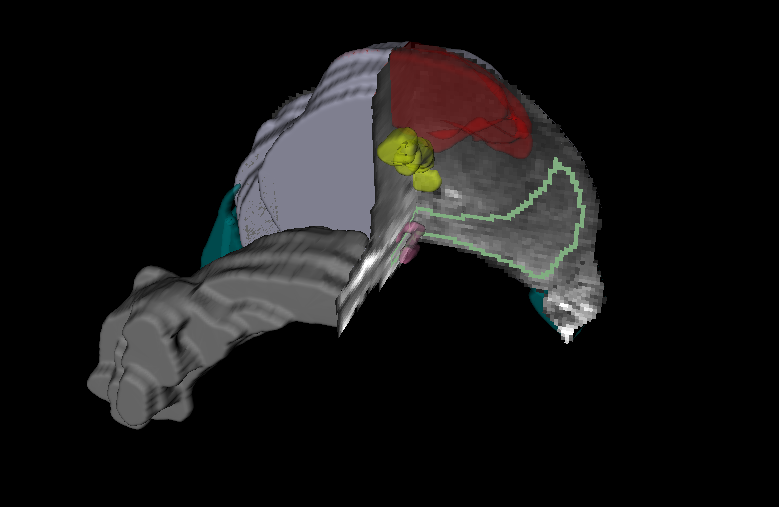
Prostate and adjacent anatomy as seen in T2w MRI. Cutoff shows the MRI intensities along with the different regions: purple-prostate capsule, light green - peripheral zone, yellow - urethra, pink - ejaculatory ducts, gray- seminal vesicles, and dark green neurovascular bundles. Also, the dominant nodule (cancer) was annotated and shown in red.

Figure by Dr. Bloch, Boston Medical Center and Drs. Mirabela Rusu and Anant Madabhushi, Center for Computational Imaging and Personalized Diagnostics, Case Western Reserve University