...
Prostate cancer T1- and T2 -weighted MRIs acquired on a 1.5 T Philips Achieva by combined surface and endorectal coil include dynamic contrast-enhanced images obtained prior to, during and after I.V. administration of 0.1 mmol/kg bodyweight of Gadolinium-DTPA. For scientific inquiries relating to the data-set please contact Drs. C. Carl Jaffe or Nicolas Bloch at carl.jaffe@bmc.org or nicolas.bloch@bmc.org.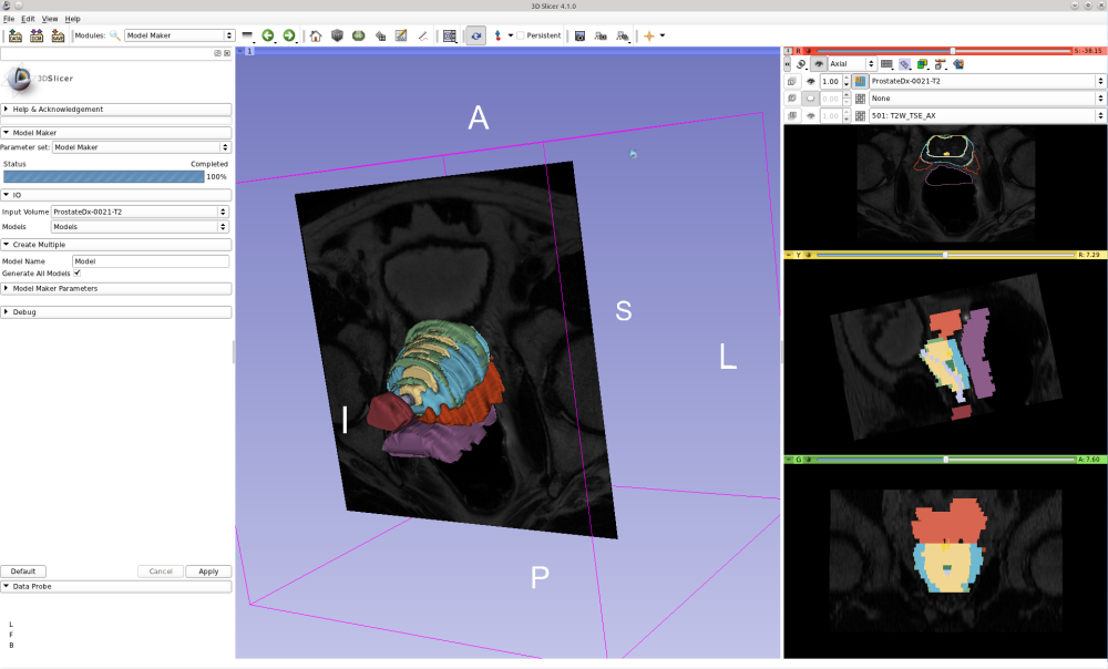
| Panel |
|---|
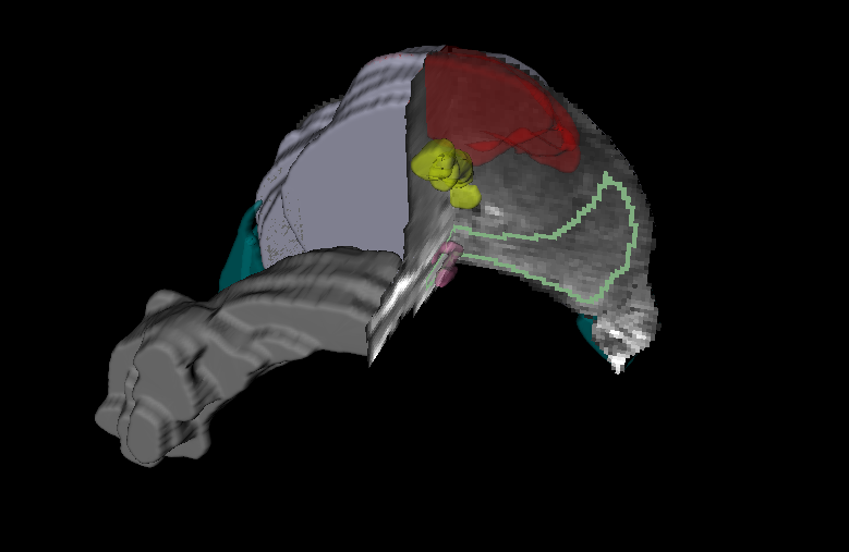
Prostate and adjacent anatomy as seen in T2w MRI. Cutoff shows the MRI intensities along with the different regions: purple-prostate capsule, light green - peripheral zone, yellow - urethra, pink - ejaculatory ducts, gray- seminal vesicles, and dark green neurovascular bundles. Also, the dominant nodule (cancer) was annotated and shown in red. Figure by Dr. Mirabela Rusu, Postdoctoral Associate, Laboratory for Computational Imaging and Bioinformatics, Rutgers State University of New Jersey. |
Supporting Documentation and metadata
...