Summary
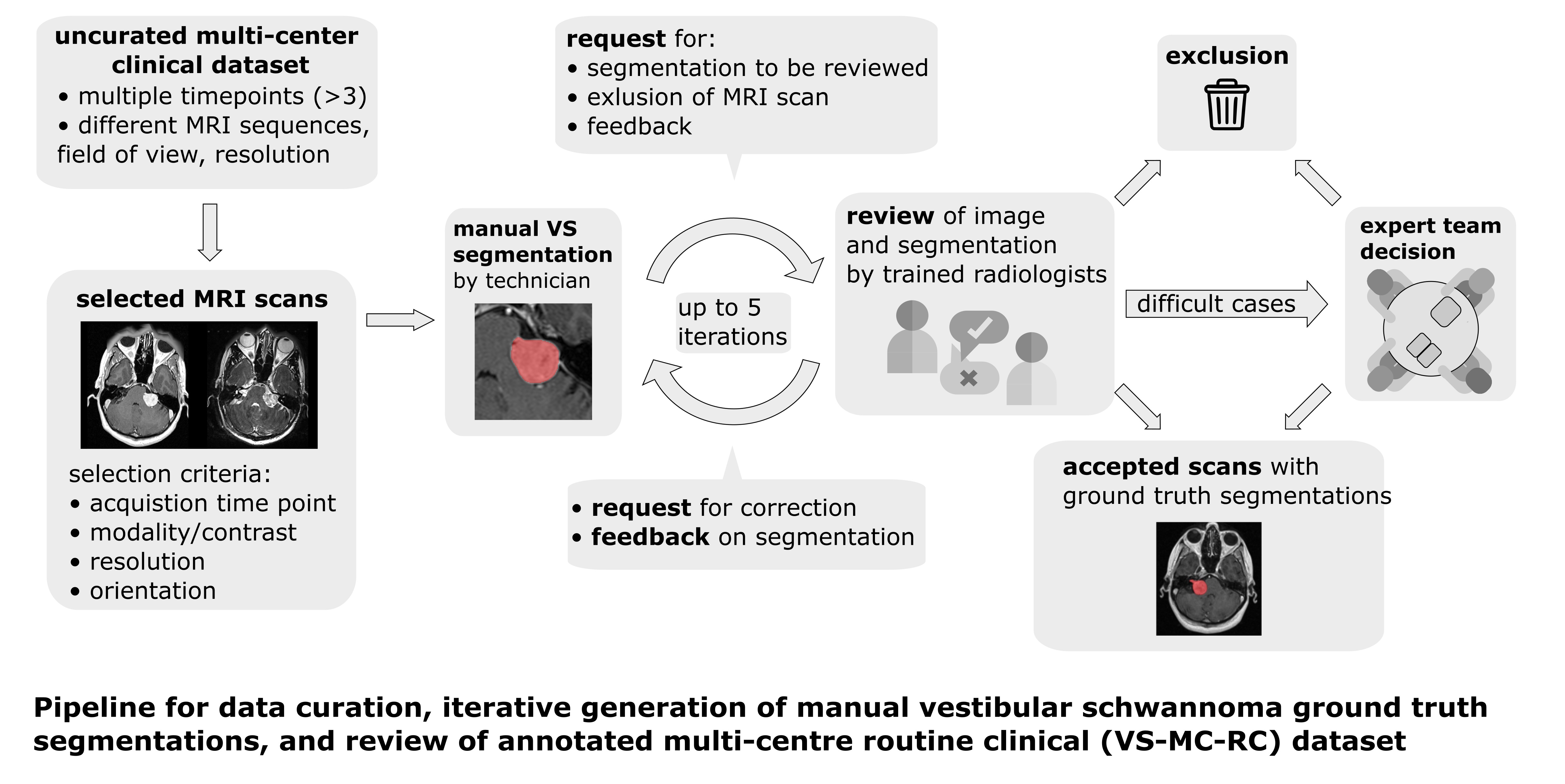
The Vestibular-Schwannoma-MC-RC (VS-MC-RC) dataset contains longitudinal MRI scans of 160 patients with a single sporadic Vestibular Schwannoma (VS) from 10 medical sites in the United Kingdom. For all patients either a T1-weighted or T2-weighted scan, or both, are available. The dataset provides binary segmentations of the VS for all scan sessions. The dataset comprises up to three time points for each patient resulting in a total of 428 timepoints and 487 3D-images. These timepoints represent the first, central, and last MRI exams that the patients underwent. Manual ground truth segmentations were obtained in an iterative process in which segmentations were: 1) produced or amended by a specialized company; and 2) reviewed by one of three trained radiologists; and 3) validated by an expert team. Using this dataset, researchers can develop and validate methods, for example, for automatic surveillance of Vestibular Schwannoma, which work robustly on images acquired at different hospitals.
Pre-processing
The MRI images are provided in DICOM format. All images were defaced using automatic affine registration or manual landmark registration with BRAINSFit and the template and face mask provided in pydeface. Segmentations are provided in NIfTI format.
Comparison to VS-SEG dataset
Compared to the Vestibular-Schwannoma-SEG (VS-SEG) dataset which was acquired with a standardized protocol for radiation treatment planning on a single scanner at a single site, the VS-MC-RC dataset aims to capture the diversity of MRI scans encountered in a routine clinical setting; it contains a wide range of scan protocols and was acquired on multiple scanners by different manufacturers. In addition, the tumour manifestation in scans of this dataset is more diverse because it includes tumours at early stages and post-operative tumour residuals rather than only tumours specifically selected for radiation therapy.
Patient overlap with VS-SEG dataset
Fifty-eight patients of the VS-MC-RC dataset were previously included in the VS-SEG dataset. These patients have a Patient ID that begins with “VS-SEG- “. Patient IDs of patients that are not part of the VS-SEG dataset begin with “VS-MC-RC-”. Please note that for most patients that are part of both datasets, the timepoints and MRI series included in one dataset are different from those included in the other dataset. However, 8 series are included in both datasets. These series are listed in the CSV file below.
Acknowledgements
We would like to acknowledge the individuals and institutions that have provided data for this collection:
This work was supported by Wellcome Trust (203145Z/16/Z, 203148/Z/16/Z, WT106882), EPSRC (NS/A000050/1, NS/A000049/1) and MRC (MC/PC/180520) funding. Additional funding was provided by Medtronic. Tom Vercauteren is also supported by a Medtronic/Royal Academy of Engineering Research Chair (RCSRF1819/7/34).
Data Access
| Data Type | Download all or Query/Filter | License |
|---|---|---|
Images (DICOM, 10 GB)* | (Download requires NBIA Data Retriever) | |
| Segmentations (NIfTI, zip) | ||
| Mapping of series in Vestibular-Schwannoma-SEG dataset (csv) |
Note: There are 124 patients currently available in TCIA search and download; the remaining 36 patients will be available post challenge in 2024.
Click the Versions tab for more info about data releases.
Detailed Description
Image Statistics | Radiology Image Statistics |
|---|---|
Modalities | MR |
Number of Patients | 124* |
Number of Studies | 301 |
Number of Series | 354 |
Number of Images | 22086 |
| Images Size (GB) | 10 |
Note: There are 124 patients currently available in TCIA search and download; the remaining 36 patients will be available post challenge in 2024.
Citations & Data Usage Policy
Users must abide by the TCIA Data Usage Policy and Restrictions. Attribution should include references to the following citations:
Data Citation
Kujawa, A., Dorent, R., Wijethilake, N., Connor, S., Thomson, S., Ivory, M., Bradford, R., Kitchen, N., Bisdas, S., Ourselin, S., Vercauteren, T., & Shapey, J. (2023). Segmentation of Vestibular Schwannoma from Magnetic Resonance Imaging: An Annotated Multi-Center Routine Clinical Dataset (Vestibular-Schwannoma-MC-RC) (Version 1) [dataset]. The Cancer Imaging Archive. https://doi.org/10.7937/HRZH-2N82
TCIA Citation
Clark, K., Vendt, B., Smith, K., Freymann, J., Kirby, J., Koppel, P., Moore, S., Phillips, S., Maffitt, D., Pringle, M., Tarbox, L., & Prior, F. (2013). The Cancer Imaging Archive (TCIA): Maintaining and Operating a Public Information Repository. In Journal of Digital Imaging (Vol. 26, Issue 6, pp. 1045–1057). Springer Science and Business Media LLC. https://doi.org/10.1007/s10278-013-9622-7
Other Publications Using This Data
TCIA maintains a list of publications which leverage TCIA data. If you have a manuscript you'd like to add please contact the TCIA Helpdesk.
Version 1 (Current): Updated 2023/08/dd
| Data Type | Download all or Query/Filter | License |
|---|---|---|
Images (DICOM, 10 GB) | (Download requires the NBIA Data Retriever) | |
| Segmentations (NIfTI, zip) | ||
| Mapping of series in Vestibular-Schwannoma-SEG dataset (csv) |
Note: There are 124 patients currently available in TCIA search and download; the remaining 36 patients will be available post challenge in 2024.