Summary
 This dataset enhances the ISPY1 data collection, with uniformly curated data, tumor annotations, and quantitative imaging features. This dataset includes a) uniformly processed scans that are harmonized to match the intensity and spatial characteristics, facilitating immediate use in computational studies, b) computationally-generated and manually-revised expert annotations of tumor regions, as well as c) a comprehensive set of quantitative imaging (also known as radiomic) features corresponding to the tumor regions.
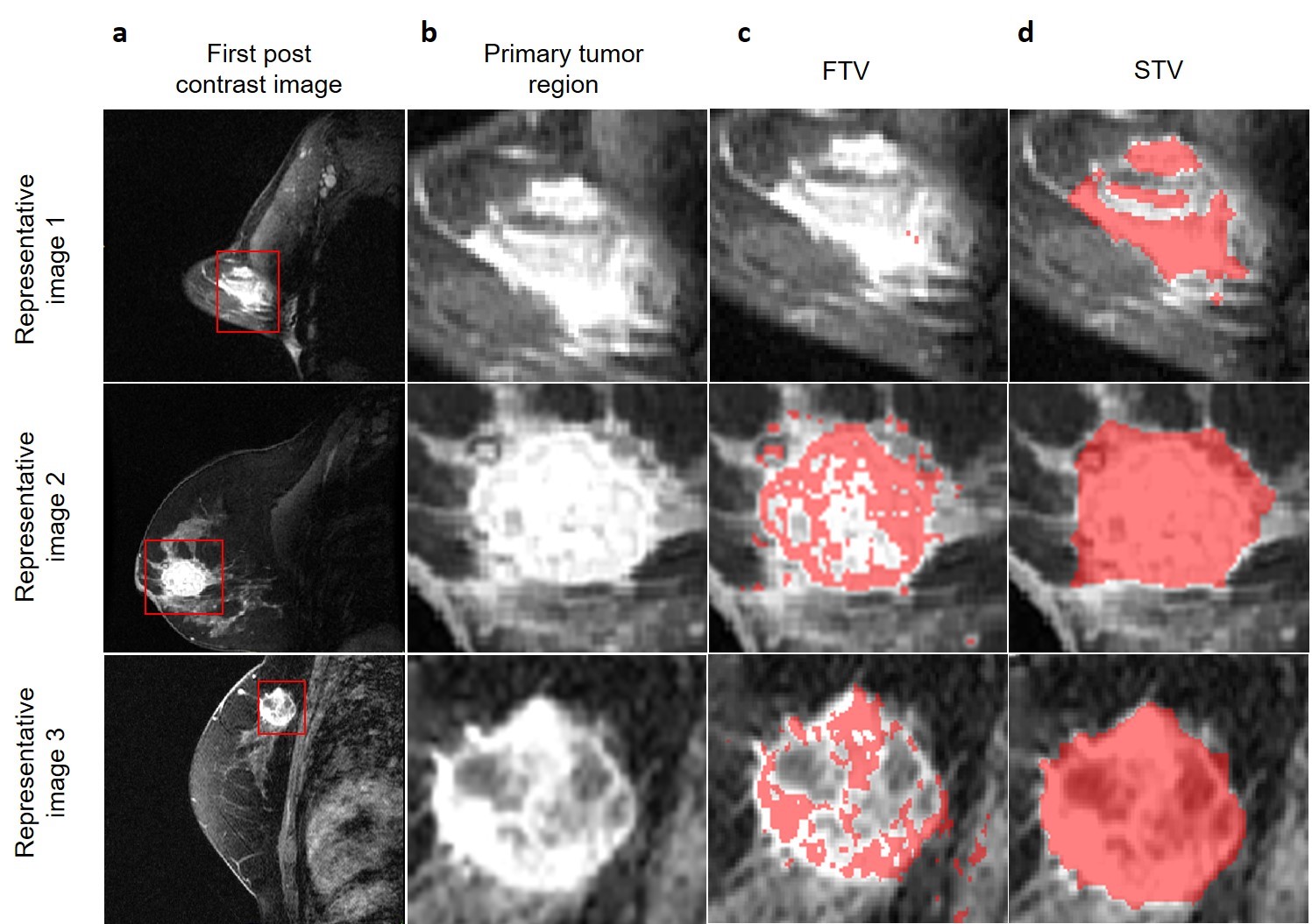
The segmentations for the ISPY1/ACRIN 6657 dataset currently hosted on TCIA’s website describe a) the tumor volume of interest (VOI) and b) functional tumor volume (FTV).
- The provided tumor VOI is a 3D rectangular box enclosing the enhancing tumor region, while including peritumoral tissue. The VOI provides a general guideline of where the tumor is located within patient anatomy, but it does not delineate tumor boundaries or shape.
- The FTV segmentations describe only enhancing voxels in the tumor, i.e., defined by peak enhancement or signal enhancement ratio criteria.
These currently provided segmentations do not include non-enhancing portions of the tumor volume, which represent a significant portion of the disease burden that needs to be studied to better understand and quantify the disease.
The segmentations in these new analysis results are for the entire 3D primary lesion, including both the enhancing and the non-enhancing tumor regions, therefore defining the structural tumor volume (STV). These STV annotations were generated by manually delineating the primary lesion volume, after confirming the location of the primary lesion from the provided VOI and FTV. The STV annotations were reviewed and approved by a board-certified, fellowship-trained breast radiologist, and are statistically significantly different from FTV.
We believe these STV annotations will allow analyses of the entire disease burden and analyses of tumor heterogeneity regarding contrast uptake, contributing to further expanding our mechanistic understanding of the disease potentially leading to improved patient management.
Acknowledgements
Research reported in this publication was partly supported by the National Cancer Institute (NCI) of the National Institutes of Health (NIH), under award numbers U01CA242871 and U24CA189523, U01CA151235, R01CA197000, and R01CA132870.
Data Access
Data Type | Download all or Query/Filter | License |
|---|---|---|
Images and Segmentations (NIfTI, 6.1 GB) | (Download and apply the IBM-Aspera-Connect plugin to your browser to retrieve this faspex package) | |
Radiomics Features (xlsx, 535 KB) | ||
README File (txt, 0.7 KB) | ||
CaPTk radiomic feature parameter (CSV, 5 KB) |
Click the Versions tab for more info about data releases.
Collections Used in this Third Party Analysis
Below is a list of the collections used in these analyses:
Source Image Data | Download or Query/Filter | License |
|---|---|---|
| ISPY1 |
Please contact help@cancerimagingarchive.net with any questions regarding usage.
Detailed Description
Image Statistics | |
|---|---|
Modalities | MR |
Number of Patients | 163 |
Number of Series/Files | 1,467 |
| Images Size (GB) | 9.8 |
Citations & Data Usage Policy
Users must abide by the TCIA Data Usage Policy and Restrictions. Attribution should include references to the following citations:
Data Citation
Chitalia, R., Pati, S., Bhalerao, M., Thakur, S., Jahani, N., Belenky, J. V., McDonald, E.S., Gibbs, J., Newitt, D., Hylton, N., Kontos, D., & Bakas, S. (2021). Expert tumor annotations and radiomic features for the ISPY1/ACRIN 6657 trial data collection [Data set]. The Cancer Imaging Archive. https://doi.org/10.7937/TCIA.XC7A-QT20
Publication Citation
Chitalia, R., Pati, S., Bhalerao, M., Thakur, S. P., Jahani, N., Belenky, V., McDonald, E. S., Gibbs, J., Newitt, D. C., Hylton, N. M., Kontos, D., & Bakas, S. (2022). Expert tumor annotations and radiomics for locally advanced breast cancer in DCE-MRI for ACRIN 6657/I-SPY1. In Scientific Data (Vol. 9, Issue 1). Springer Science and Business Media LLC. https://doi.org/10.1038/s41597-022-01555-4
TCIA Citation
Clark K, Vendt B, Smith K, Freymann J, Kirby J, Koppel P, Moore S, Phillips S, Maffitt D, Pringle M, Tarbox L, Prior F. The Cancer Imaging Archive (TCIA): Maintaining and Operating a Public Information Repository, Journal of Digital Imaging, Volume 26, Number 6, December, 2013, pp 1045-1057. DOI: 10.1007/s10278-013-9622-7
Other Publications Using This Data
TCIA maintains a list of publications which leverage TCIA data. If you have a manuscript you'd like to add please contact the TCIA Helpdesk.
Version 1 (Current): 2022/06/01
| Data Type | Download all or Query/Filter |
|---|---|
| Images and Segmentations (NIfTI, 6.1 GB) | (Download requires IBM-Aspera-Connect plugin to your browser) |
| Radiomics Features (xlsx) | |
| README File (txt) |