Summary
| Excerpt |
|---|
This collection consists of 4 image sets generated at University of Iowa and University of Washington. It was originally contributed for use in a positron emission tomography (PET) phantom scans originally utilized by the Quantitative Imaging Network (QIN) PET Segmentation Challenge . |
HIGH Statistics (HS) 30 minute Scan with HIGH Contrast (HC) [9.77:1]

HIGH Statistics (HS) 30 minute Scan with LOW Contrast (LC) [4.88:1]

10 x LOW Statistics (LS) 3 minute Scan with HIGH Contrast (HC) [9.77:1]

10 x LOW Statistics (LS) 3 minute Scan with LOW Contrast (LC) [4.88:1]

to assess the variability of segmentations and subsequently derived quantitative analysis results on phantom PET scans with known ground truth. |
The phantom was provided by Dr. Sunderland at the University of Iowa (supported by grant R01CA169072 - Harmonized PET Reconstructions for Cancer Clinical Trials) and is based on the NEMA IEC Body Phantom SetTM (Model PET/IEC-BODY/P) with a set of 6 custom made (via rapid prototyping) spheres & ellipses (Fig. 1). The phantom was scanned at two QIN sites (University of Iowa and University of Washington) with different scanners, following a protocol that yields four image sets (Fig. 2) per site. The DICOM PET images are organized as shown in Fig. 3.
Please send questions regarding the QIN PET to qin_pet_challenge@iibi.uiowa.edu.

Fig. 1. Phantom used in the QIN PET challenge.
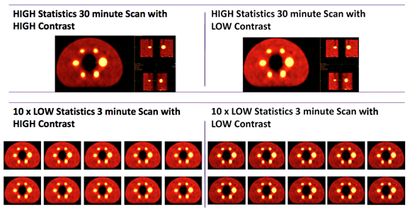
Fig. 2. Overview of image sets.
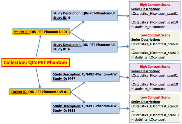
Fig. 3. Organization of DICOM PET scans.
About the NCI QIN
The mission of the QIN is to improve the role of quantitative imaging for clinical decision making in oncology by developing and validating data acquisition, analysis methods, and tools to tailor treatment for individual patients and predict or monitor the response to drug or radiation therapy. More information is available on the Quantitative Imaging Network Collections page. Interested investigators can apply to the QIN at: Quantitative Imaging for Evaluation of Responses to Cancer Therapies (U01) PAR-11-150.
Data Access
Imaging Data
Collection Statistics | Updated 2014/09/04 |
|---|---|
Modalities | PT |
Number of Patients | 2 |
Number of Studies | 4 |
Number of Series | 44 |
Number of Images | 2,816 |
| Images Size (GB) | 0.246 |
If you are unsure how to download this collection, please view Searching by Collection or refer to our TCIA's User's Guide for more detailed instructions on using the site.
Shared Lists
- None available at this time.