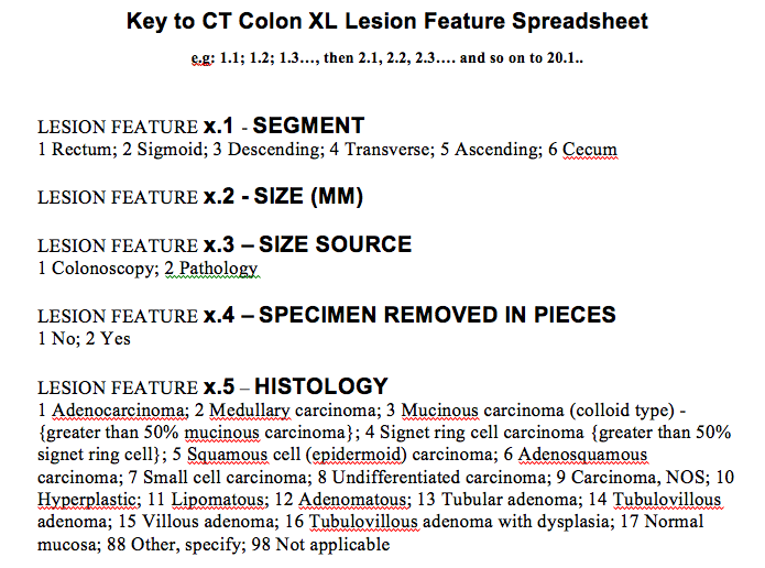
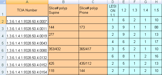
Versions Compared

Key

  • This line was added.
  • This line was removed.
  • Formatting was changed.

Summary

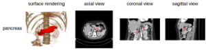
In 2004 when presenting the NCI Executive Committee the ACRIN proposal to conduct the CTC trial, a case was made that publicly accessible image data sharing would offer a valuable research asset to a wide image processing research community. Adding to the many merits of that proposal, the data-sharing component was strongly endorsed. ACRIN completed the trial expeditiously and its results were published in NEJM in fall 2008 to wide interest. ACRIN has graciously allowed the wider research community access to a portion of the data from that trial here on TCIA, including spreadsheets identifying positive and negative polyp cases. The complete ACRIN protocol description can be found at 

The National Institutes of Health Clinical Center performed 82 abdominal contrast enhanced 3D CT scans (~70 seconds after intravenous contrast injection in portal-venous) from 53 male and 27 female subjects.  Seventeen of the subjects are healthy kidney donors scanned prior to nephrectomy.  The remaining 65 patients were selected by a radiologist from patients who neither had major abdominal pathologies nor pancreatic cancer lesions.  Subjects' ages range from 18 to 76 years with a mean age of 46.8 ± 16.7. The CT scans have resolutions of 512x512 pixels with varying pixel sizes and slice thickness between 1.5 − 2.5 mm, acquired on Philips and Siemens MDCT scanners (120 kVp tube voltage).

A medical student manually performed slice-by-slice segmentations of the pancreas as ground-truth and these were verified/modified by an experienced radiologist.

Data Example


Image Added

Note

The DICOM files were created from anonymized volumetric images (Analyze and NifTI) using this from ITK: http://www.

acrin

itk.org/

TabID

Doxygen/

151/Default.aspx

html/Examples_2IO_2ImageReadDicomSeriesWrite_8cxx-example.html.


Localtab Group
Localtab
activetrue
titleData Access

Data Access

Choosing the Download option will provide you with a file to launch the TCIA Download Manager to download the entire collection. If you want to browse or filter the data to select only specific scans/studies please use the Search By Collection option.

Data TypeDownload all or Query/Filter
Images (DICOM, 46210.6GB2 GB)Polyp Descriptions - No polyp found (XLS) Image Removed
Polyp Descriptions - Large 10mm (XLS)Image Removed
Polyp Descriptions - 6 to 9mm (XLS)Image Removed
Image Modified

Click the Versions tab for more info about data releases.

Localtab
titleDetailed Description

Detailed Description

Collection Statistics


Modalities

CT

Number of Patients

825 

Number of Studies

836 

Number of Series

3,451 

Number of Images

941,771 

Image Size (GB)462.6
There are presently 825 cases in this collection with XLS sheets that provide polyp descriptions and their location within the colon segments.  To link the XLS polyp tables with the DICOM image studies in TCIA you should understand that some cases in the TCIA are identified by long numbers with the last 4 digits after the last decimal point (e.g.: NCIA study number "1.3.6.1.4.1.9328.50.4.0040" referred to as case "40"). In addition there are a fewer number of additional positive cases that begin their identification number with 'CTC' (e.g.: CTC-5401799343
)

Three related XLS spreadsheets are in this release. 

TCIA CTC large
10
mm polyps.xls - Contains the case numbers for 35 cases (out of the 825 total TCIA cases) where at least one 10mm or larger size polyp was found.  Individual cases may have several (up to 20) polyps of different sizes listed on a particular XLS row as "LESION 1
.
x,
2
.x,3.x etc. – see "feature key" below).
  • TCIA CTC 6 to 9 mm polyps.xls - Contains 69 cases with smaller size polyps.
  • TCIA CTC no polyp found.xls - Contains 243 cases that were recorded as free of polyps by both CTC and optical techniques.
  • Thus in this CT Colonography collection you will be able to download the prone and supine DICOM images from OC same-day validated 243 negative cases, 69 cases with 6 to 9 mm polyps, and 35 cases which have at least one > 10 mm polyp and their histological type. Below is the key for deciphering the features in the spreadsheet.

    Image Removed

    WARNING: NCI cannot assure archive users of error-free validity of the XL polyp location data since NCI did not itself perform the clinical study or its analysis.

    You will note that two XLS files with positive findings have multiple columns descriptors of individual polyp lesions listed as in the table below.  The meaning of the colored columns labeled "LESION 1.1...1.2...1.3...1.4, etc" is explained in the attached key-code ".tiff" file entitled "Polyp description key table.tiff"). Some CT scan slice numbers where the polyps were found are provided, but unfortunately the table may not have complete slice number information – you'll just have to do the best you can with the data NCI was given.

    Image Removed


    Localtab
    titleCitations & Data Usage Policy

    Citations & Data Usage Policy 

    This collection is freely available to browse, download, and use for commercial, scientific and educational purposes as outlined in the Creative Commons Attribution 3.0 Unported License.  See TCIA's Data Usage Policies and Restrictions for additional details. Questions may be directed to help@cancerimagingarchive.net.

    Please be sure to include the following citations in your work if you use this data set:

    Info
    titleCT Colonography Citation

    The Cancer Imaging Archive Team. Data From CT_Colonography. doi:10.7937/K9/TCIA.2015.NWTESAY1


    Info
    titlePublication Citation

    C.D. Johnson, MD, MMM,M-H. Chen, PhD, A.Y. Toledano, ScD, J.P. Heiken, MD, A. Dachman, MD, M.D. Kuo, MD, C. Menias, MD, B. Siewert, MD, J.I. Cheema, MD, R.G. Obregon, MD, J.L. Fidler, MD, P. Zimmerman, MD, K.M. Horton, MD, K. Coakley, MD, R.B. Iyer, MD, A.K. Hara, MD, R.A. Halvorsen, Jr., MD, G. Casola, MD, J. Yee, MD, B. A. Herman, SM, L.J. Burgart, MD, and P.J. Limburg, MD, MPH. Accuracy of CT Colonography for Detection of Large Adenomas and Cancers. N Engl J Med. 2008 Sep 18; 359(12): 1207–1217. doi:  10.1056/NEJMoa0800996. (Roth HR, Lu L, Farag A, Shin H-C, Liu J, Turkbey EB, Summers RM. DeepOrgan: Multi-level Deep Convolutional Networks for Automated Pancreas Segmentation. N. Navab et al. (Eds.): MICCAI 2015, Part I, LNCS 9349, pp. 556–564, 2015.  (paper)

    Info
    titleTCIA Citation

    Clark K, Vendt B, Smith K, Freymann J, Kirby J, Koppel P, Moore S, Phillips S, Maffitt D, Pringle M, Tarbox L, Prior F. The Cancer Imaging Archive (TCIA): Maintaining and Operating a Public Information Repository, Journal of Digital Imaging, Volume 26, Number 6, December, 2013, pp 1045-1057. (paper)

    Other Publications Using This Data

    See the CT Colonography section on our Publications page for other work leveraging this collection. If you have a publication you'd like to add please contact the TCIA Helpdesk.

    Localtab
    titleVersions

    Version 1 (Current): Updated

    2013/11/15

    20151229

    Data TypeDownload all or Query/Filter
    Images (DICOM, 46210.6GB2 GB)Polyp Descriptions - No polyp found (XLS) Image Removed
    Polyp Descriptions - Large 10mm (XLS)Image Removed
    Polyp Descriptions - 6 to 9mm (XLS)Image Removed
    Image Modified