...
| Excerpt |
|---|
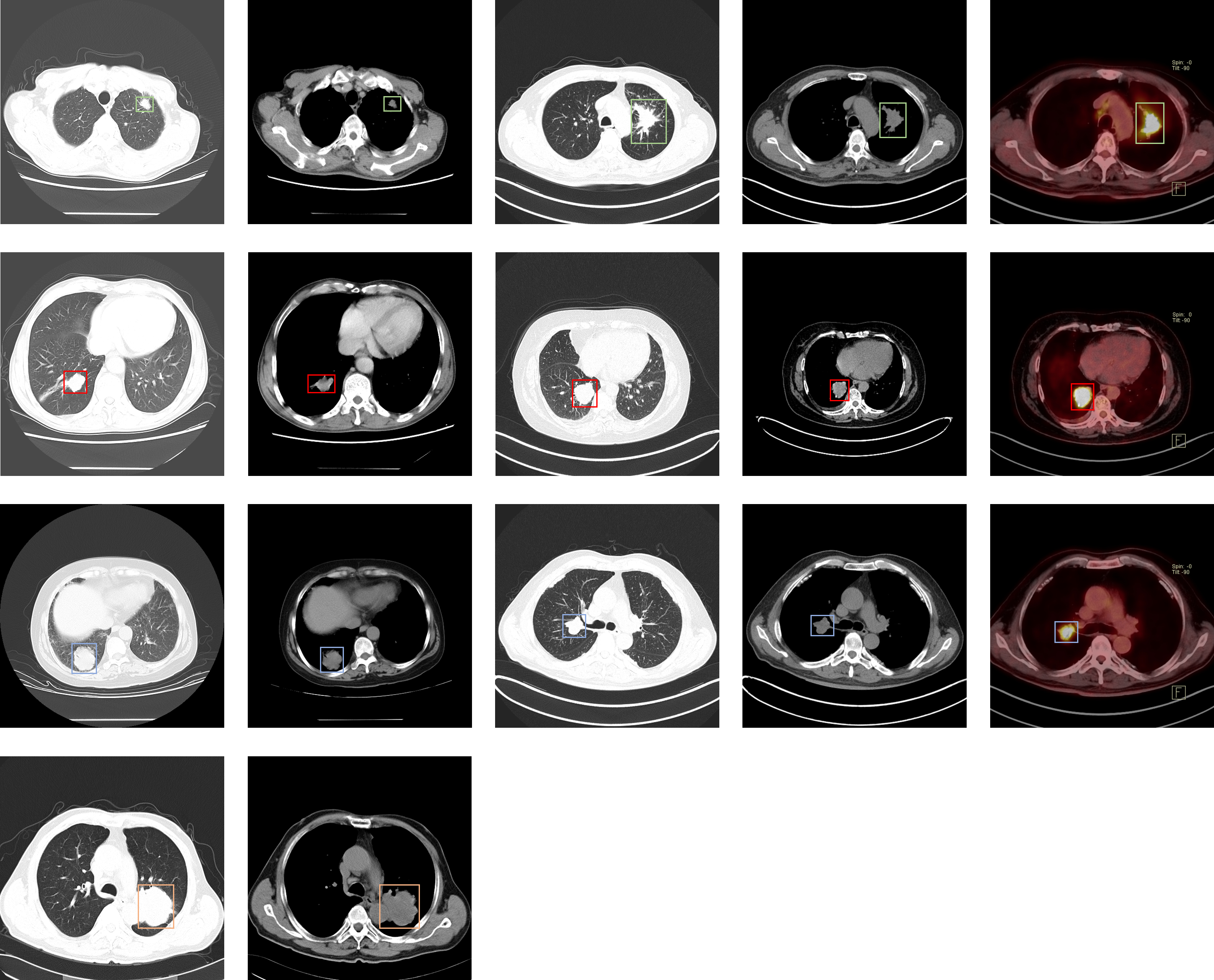 This dataset consists of CT and PET-CT DICOM images of lung cancer subjects with XML Annotation files that indicate tumor location. The images were retrospectively acquired from patients with suspicion of lung cancer, and who underwent standard-of-care lung biopsy and PET/CT. The cases were confirmed by pathological diagnosis. The images were analyzed on the mediastinum (window width, 350 HU; level, 40 HU) and lung (window width, 1,400 HU; level, –700 HU) settings. The reconstructions were made in 2mm-slice-thick and lung settings. The CT slice interval varies from 0.625 mm to 5 mm. Scanning mode includes plain and contrast and 3D reconstruction. The location of the tumors were each tumor was provided by five chest radiologists and two deep learning researchers in order to make this dataset a useful tool and resource for developing algorithms for medical diagnosis. The image annotations are saved as XML files in PASCAL VOC format, which can be parsed using the PASCAL Development Toolkit: https://pypi.org/project/pascal-voc-tools/. |
...