Versions Compared

Key

  • This line was added.
  • This line was removed.
  • Formatting was changed.

Summary

Excerpt

This These retrospective NIfTI image dataset datasets consists of unenhanced chest CTs

  • First dataset - from 632 patients with COVID-19 infections at initial point of care, and
  • Second dataset - a second set of 121 CTs from 29 patients with COVID-19 infections with serial / sequential CTs.

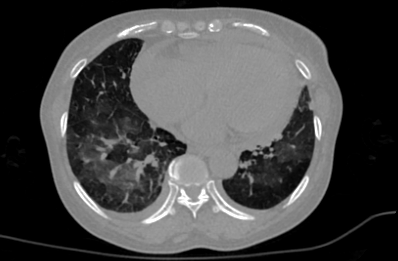
The initial images for both datasets were acquired at acquired at the point of care in an outbreak setting from setting from patients with Reverse Transcription Polymerase Chain Reaction (RT-PCR) confirmation for the presence of SARS-CoV-2.

Patients presented to a health care setting with a combination of symptoms, exposure to an infected patient, or travel history to an outbreak region. All patients had a positive RT-PCR for SARS-CoV-2 from a sample obtained within 1 day of the initial CT.


CT exams were performed without intravenous contrast and with a soft tissue reconstruction algorithm.  The DICOM images were subsequently converted into NIfTI format

. The second dataset also had other follow up CTs, in addition to the initial point of care CT.

A multidisciplinary team trained several models using portions of this TCIA data setthe first dataset, along with additional CTs and manually annotated images from other sources. A classification model derived in part from this data the first dataset is described in a Nature Communications manuscript at:  https://doi.org/10.1038/s41467-020-17971-2The NVIDIA-related frameworks and models specific to this publication are available at no cost as part of the NVIDIA Clara Train SDK at https://ngc.nvidia.com/catalog/containers/nvidia:clara:ai-covid-19. This includes both inference-based pipelines for evaluation, as well as model weights for further training or fine tuning in outside institutions. The second data set of 121 serial / sequential CTs in 29 patients is reported in a Scientific Reports manuscript at  https://doi.org/10.1038/s41598-021-85694-5In addition, a web-based version of this model (for research use only) with drag-and-drop functionality for evaluating individual scans can be found at https://marketplace.arterys.com/model/nvidiacovidCT.  Uploading a CT yields a return email that contains results. 

...

Localtab Group


Localtab
activetrue
titleData Access

Data Access

Data TypeDownload all or Query/Filter

Images (NIfTI, 12.71 GB)

First dataset

Tcia button generator
urlhttps://faspex.cancerimagingarchive.net/aspera/faspex/external_deliveries/44?passcode=7f03b74391b2da371d9e393255847aa60027f6c0

Images (NIFTI, )

Second dataset

Tcia button generator
urlhttps://faspex.cancerimagingarchive.net/aspera/faspex/external_deliveries/134?passcode=c6849b3481a1b240daebc7e3c50e1e4f7e3fa424

Click the Versions tab for more info about data releases.

Please contact help@cancerimagingarchive.net  with any questions regarding usage.


Localtab
titleDetailed Description

Detailed Description

Image Statistics


Modalities

CT

Number of Patients

632661

Number of Studies

650771
Images Size (GB)1214.717


Localtab
titleCitations & Data Usage Policy

Citations & Data Usage Policy

Tcia license 4 international

Info
titleData Citation

An P, Xu S, Harmon SA, Turkbey EB, Sanford TH, Amalou A, Kassin M, Varble N, Blain M, Anderson V, Patella F, Carrafiello G, Turkbey BT, Wood BJ (2020). CT Images in Covid-19 [Data set]. The Cancer Imaging Archive. DOI: https://doi.org/10.7937/tcia.2020.gqry-nc81


Info
titlePublication Citation

Link to publication below contains AI model that was only partly derived from this data, but also from other data, not present here on TCIA.

Harmon SA, Sanford TH, Xu S, Turkbey EB, Roth H, Xu Z, Yang D, Myronenko A, Anderson V, Amalou A, Blain M, Kassin M, Long D, Varble N, Walker SM, Bagci U, Ierardi AM, Stellato E, Plensich GG, Franceschelli G, Girlando C, Irmici G, Labella D, Hammoud D, Malayeri A, Jones E, Summer RM, Choyke PL, Xu D, Flores M, Tamura K, Obinata H, Mori H, Patella F, Cariati M, Carrafiello G, An P, Wood BJ, Turkbey B (2020). Artificial intelligence for the detection of COVID-19 pneumonia on chest CT using multinational datasets. Nature Communications. DOI: https://doi.org/10.1038/s41467-020-17971-2


Info
titleAcknowledgement

The Multi-national NIH Consortium for CT AI in COVID-19.


Info
titleTCIA Citation

Clark K, Vendt B, Smith K, Freymann J, Kirby J, Koppel P, Moore S, Phillips S, Maffitt D, Pringle M, Tarbox L, Prior F. The Cancer Imaging Archive (TCIA): Maintaining and Operating a Public Information Repository, Journal of Digital Imaging, Volume 26, Number 6, December, 2013, pp 1045-1057. DOI: 10.1007/s10278-013-9622-7.

Other Publications Using This Data

TCIA maintains a list of publications which leverage TCIA data. If you have a manuscript you'd like to add please contact the TCIA Helpdesk.


Localtab
titleVersions

Version

1 (Current)

2 (Current): Updated 2021/05/21

Data TypeDownload all or Query/Filter

Images (NIfTI, 12.71 GB)

First dataset

Tcia button generator
urlhttps://faspex.cancerimagingarchive.net/aspera/faspex/external_deliveries/44?passcode=7f03b74391b2da371d9e393255847aa60027f6c0

Images (NIFTI, 2 GB)

Second dataset

Tcia button generator
urlhttps://faspex.cancerimagingarchive.net/aspera/faspex/external_deliveries/134?passcode=c6849b3481a1b240daebc7e3c50e1e4f7e3fa424

Added second dataset, 29 patients/121 CT images.

Version 1: Updated 2020/08/31

Data TypeDownload all or Query/Filter

Images (NIfTI, 12.71 GB)

Tcia button generator
urlhttps://faspex.cancerimagingarchive.net/aspera/faspex/external_deliveries/44?passcode=7f03b74391b2da371d9e393255847aa60027f6c0


...