Summary
| Excerpt |
|---|
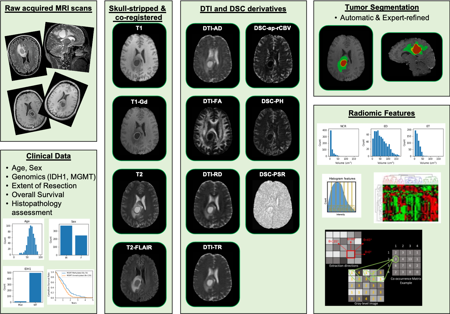
This collection comprises multi-parametric magnetic resonance imaging (mpMRI) scans for de novo Glioblastoma (GBM) patients from the Hospital of the University of Pennsylvania, coupled with patient demographics, clinical outcome (e.g., overall survival, genomic information, tumor progression), as well as computer-aided and manually-corrected segmentation labels of multiple histologically distinct tumor sub-regions, computer-aided and manually-corrected segmentations of the whole brain, a rich panel of radiomic features along with their corresponding co-registered mpMRI volumes in NIfTI format. Scans were initially skull-stripped and co-registered, before their tumor segmentation labels were produced by an automated computational method. These segmentation labels were revised and any label misclassifications were manually corrected/approved by expert board-certified neuroradiologists. The final labels were used to extract a rich panel of imaging features, including intensity, volumetric, morphologic, histogram-based and textural parameters. The segmentation labels enable quantitative computational and clinical studies without the need to repeat manual annotations whilst allowing for comparison across studies. They can also serve as a set of manually-annotated gold standard labels for performance evaluation in computational challenges. The provided panel of radiomic features may facilitate research integrative of the molecular characterization offered, and hence allow associations with molecular markers (radiogenomic biomarker research), clinical outcomes, treatment responses and other endpoints, by researchers without sufficient computational background to extract such features. |
Acknowledgements
We would like to acknowledge the individuals and institutions that have provided data for this collection:
...
| Localtab Group |
|---|
| Localtab |
|---|
| active | true |
|---|
| title | Data Access |
|---|
| Data AccessClick the Download button to save a ".tcia" manifest file to your computer, which you must open with the NBIA Data Retriever. Click the Search button to open our Data Portal, where you can browse the data collection and/or download a subset of its contents. | Data Type | Download all or Query/Filter |
|---|
Images (DICOM, XX.X GB)
| | | NIfTI Files | | | Clinical Data (CSV) | |
Click the Versions tab for more info about data releases. Please contact help@cancerimagingarchive.net with any questions regarding usage. |
| Localtab |
|---|
| title | Detailed Description |
|---|
| Detailed DescriptionImage Statistics |
|
|---|
Modalities | MR | Number of Patients | 630 | Number of Studies | 3299 | Number of Series | 3679 | Number of Images | 826,782 | | Images Size (GB) | 139.3 |
|
| Localtab |
|---|
| title | Citations & Data Usage Policy |
|---|
| Citations & Data Usage Policy| Tcia license 4 international |
|---|
This is a limited access data set and is only available to members of UPENN. If you are a member of UPENN and would like to request access, please submit a CCP proposal to the Coordinating Committee. Upon receiving access you may only use it for the purposes outlined in your proposal. Questions may be directed to help@cancerimagingarchive.net.
| Info |
|---|
| title | Publication Citation |
|---|
| We ask on the proposal form if they have ONE traditional publication they'd like users to cite. |
| Info |
|---|
| Clark, K., Vendt, B., Smith, K., Freymann, J., Kirby, J., Koppel, P., Moore, S., Phillips, S., Maffitt, D., Pringle, M., Tarbox, L., & Prior, F. (2013). The Cancer Imaging Archive (TCIA): Maintaining and Operating a Public Information Repository. Journal of Digital Imaging, 26(6), 1045–1057. https://doi.org/10.1007/s10278-013-9622-7 |
Other Publications Using This DataTCIA maintains a list of publications which leverage TCIA data. If you have a manuscript you'd like to add please contact the TCIA Helpdesk. |
| Localtab |
|---|
| Version 1 (Current): 2021/00/00| Data Type | Download all or Query/Filter |
|---|
| Images (DICOM, xx.x GB) | | | NIfTI Files | | | Clinical Data (CSV) | |
|
|
...