| Localtab |
|---|
| active | true |
|---|
| title | Data Access |
|---|
| Data AccessChoosing the Download option will provide you with a file to launch the TCIA Download Manager to download the entire collection. If you want to browse or filter the data to select only specific scans/studies please use the Search By Collection option.
| Data Type | Download all or Query/Filter | |
|---|
| Images (DICOM, |
4626GB) Image Removed Image Removed Image Removed Image Removed
| | Polyp Descriptions - Large 10mm (XLS) | Image Removed | | Polyp Descriptions - 6 to 9mm (XLS) | Image Removed | Polyp Descriptions - No polyp found (XLS) | | 3 GB) |
| Tcia button generator |
|---|
| url | https://wiki.cancerimagingarchive.net/download/attachments/22514040/Pancreas-CT%2020200910.tcia?version=1&modificationDate=1599754273288&api=v2 |
|---|
|
|
| Tcia button generator |
|---|
| label | Search |
|---|
| url | https://www.cancerimagingarchive.net/nbia-search/?CollectionCriteria=Pancreas-CT |
|---|
|
|
(Download requires NBIA Data Retriever) | | | Manual Annotations (zip, 975 kB) |
| Tcia button generator |
|---|
| url | https://wiki.cancerimagingarchive.net/download/attachments/22514040/TCIA_pancreas_labels-02-05-2017.zip?version=2 |
|---|
|
|
| |
Image Removed
Click the Versions tab for more info about data releases. | Nci_crdc additional resources |
|---|
|
| Localtab |
|---|
| title | Detailed Description |
|---|
| Detailed Description
Patients8258363,451941771| 462.6 | There are presently 825 cases in this collection with XLS sheets that provide polyp descriptions and their location within the colon segments. To link the XLS polyp tables with the DICOM image studies in TCIA you should understand that some cases in the TCIA are identified by long numbers with the last 4 digits after the last decimal point (e.g.: NCIA study number "1.3.6.1.4.1.9328.50.4.0040" referred to as case "40"). In addition there are a fewer number of additional positive cases that begin their identification number with 'CTC' (e.g.: CTC-5401799343) Three related XLS spreadsheets are in this release. - TCIA CTC large 10 mm polyps.xls - Contains the case numbers for 35 cases (out of the 825 total TCIA cases) where at least one 10mm or larger size polyp was found. Individual cases may have several (up to 20) polyps of different sizes listed on a particular XLS row as "LESION 1.x, 2.x,3.x etc. – see "feature key" below).
- TCIA CTC 6 to 9 mm polyps.xls - Contains 69 cases with smaller size polyps.
- TCIA CTC no polyp found.xls - Contains 243 cases that were recorded as free of polyps by both CTC and optical techniques.
Thus in this CT Colonography collection you will be able to download the prone and supine DICOM images from OC same-day validated 243 negative cases, 69 cases with 6 to 9 mm polyps, and 35 cases which have at least one > 10 mm polyp and their histological type. Below is the key for deciphering the features in the spreadsheet. 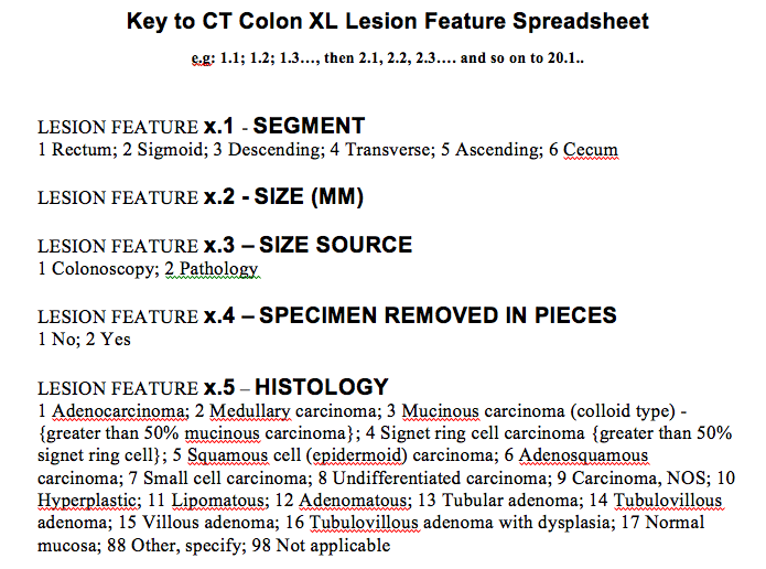 Image Removed Image Removed
WARNING: NCI cannot assure archive users of error-free validity of the XL polyp location data since NCI did not itself perform the clinical study or its analysis. You will note that two XLS files with positive findings have multiple columns descriptors of individual polyp lesions listed as in the table below. The meaning of the colored columns labeled "LESION 1.1...1.2...1.3...1.4, etc" is explained in the attached key-code ".tiff" file entitled "Polyp description key table.tiff"). Some CT scan slice numbers where the polyps were found are provided, but unfortunately the table may not have complete slice number information – you'll just have to do the best you can with the data NCI was given. 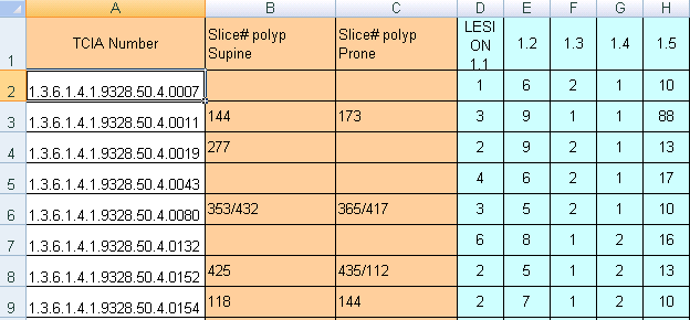 Image Removed Image Removed
Data Example
 Image Added Image Added
NoteThe DICOM files were created from anonymized volumetric images (Analyze and NifTI) using this from ITK: http://www.itk.org/Doxygen/html/Examples_2IO_2ImageReadDicomSeriesWrite_8cxx-example.html .
|
| Localtab |
|---|
| title | Citations & Data Usage Policy |
|---|
| Citations & Data Usage Policy This collection is freely available to browse, download, and use for commercial, scientific and educational purposes as outlined in the Creative Commons Attribution 3.0 Unported License. See TCIA's Data Usage Policies and Restrictions for additional details. Questions may be directed to help@cancerimagingarchive.net. Please be sure to include the following citations in your work if you use this data set: | Info |
|---|
| title | CT Colonography Citation |
|---|
| The Cancer Imaging Archive Team. Data From CT_Colonography. doi:10.7937/K9/TCIA.2015.NWTESAY1 |
| Tcia limited license policy |
|---|
| Info |
|---|
| Roth, H., Farag, A., Turkbey, E. B., Lu, L., Liu, J., & Summers, R. M. (2016). Data From Pancreas-CT (Version 2) [Data set]. The Cancer Imaging Archive. https://doi.org/10.7937/K9/TCIA.2016.tNB1kqBU |
| Info |
|---|
| title | Publication Citation |
|---|
| Roth HR, Lu L, Farag A, Shin H-C, Liu J, Turkbey EB, Summers RM. DeepOrgan: Multi-level Deep Convolutional Networks for Automated Pancreas Segmentation. N. Navab et al. (Eds.): MICCAI 2015, Part I, LNCS 9349, pp. 556–564, 2015. (arXiv link) https://doi.org/10.1007/978-3-319-24553-9_68 | | Info |
|---|
| title | Publication Citation |
|---|
| C.D. Johnson, MD, MMM,M-H. Chen, PhD, A.Y. Toledano, ScD, J.P. Heiken, MD, A. Dachman, MD, M.D. Kuo, MD, C. Menias, MD, B. Siewert, MD, J.I. Cheema, MD, R.G. Obregon, MD, J.L. Fidler, MD, P. Zimmerman, MD, K.M. Horton, MD, K. Coakley, MD, R.B. Iyer, MD, A.K. Hara, MD, R.A. Halvorsen, Jr., MD, G. Casola, MD, J. Yee, MD, B. A. Herman, SM, L.J. Burgart, MD, and P.J. Limburg, MD, MPH. Accuracy of CT Colonography for Detection of Large Adenomas and Cancers. N Engl J Med. 2008 Sep 18; 359(12): 1207–1217. doi: 10.1056/NEJMoa0800996. (paper) |
| Info |
|---|
| Clark K, Vendt B, Smith K, Freymann J, Kirby J, Koppel P, Moore S, Phillips S, Maffitt D, Pringle M, Tarbox L, Prior F. The Cancer Imaging Archive (TCIA): Maintaining and Operating a Public Information Repository, Journal of Digital Imaging, Volume 26, Number 6, December, 2013, pp 1045-1057. (paper). DOI: https://doi.org/10.1007/s10278-013-9622-7 |
Other Publications Using This DataSee the CT Colonography section on our Publications page for other work leveraging this collection. TCIA maintains a list of publications that leverage TCIA data. If you have a publication manuscript you'd like to add please contact the TCIA Helpdesk. Below is a list of such publications using this Collection: - Gibson, E., Giganti, F., Hu, Y., Bonmati, E., Bandula, S., Gurusamy, K., . . . Barratt, D. C. (2017). Towards Image-Guided Pancreas and Biliary Endoscopy: Automatic Multi-organ Segmentation on Abdominal CT with Dense Dilated Networks. Paper presented at the International Conference on Medical Image Computing and Computer-Assisted Intervention.
- Greenspan, H., van Ginneken, B., & Summers, R. M. (2016). Guest Editorial Deep Learning in Medical Imaging: Overview and Future Promise of an Exciting New Technique. IEEE Transactions on Medical Imaging, 35(5), 1153-1159. doi:10.1109/TMI.2016.2553401
- Shi, H., Lu, L., Yin, M., Zhong, C., & Yang, F. (2023). Joint few-shot registration and segmentation self-training of 3D medical images. Biomedical Signal Processing and Control, 80. doi:https://doi.org/10.1016/j.bspc.2022.104294
|
| Localtab |
|---|
| Version 1 2 (Current): Updated 20132020/1109/1510 |
|