Summary
| Redirect |
|---|
| delay | 5 |
|---|
| location | https://www.cancerimagingarchive.net/collection/prostatex/ |
|---|
|
| Info |
|---|
| title | PROSTATEx has been superseded by PI-CAI |
|---|
|
The ProstateX |
Summary
| Excerpt |
|---|
SPIE, along with the support of the American Association of Physicists in Medicine (AAPM) and the National Cancer Institute (NCI), will conduct a “Grand Challenge” on dataset (both training and testing cases) have been included in the PI-CAI Public Training and Development dataset. As such, ProstateX as a benchmark has been deprecated and is superseded by the PI-CAI challenge. PI-CAI is an all-new grand challenge, with over 10,000 carefully-curated prostate MRI exams to validate modern AI algorithms and estimate radiologists' performance at clinically significant prostate cancer detection and diagnosis. Key aspects of the study design have been established in conjunction with an international, multi-disciplinary scientific advisory board (16 experts in prostate AI, radiology and urology) - to unify and standardize present-day guidelines, and to ensure meaningful validation of prostate-AI towards clinical translation. Please refer to https://pi-cai.grand-challenge.org for more information. |
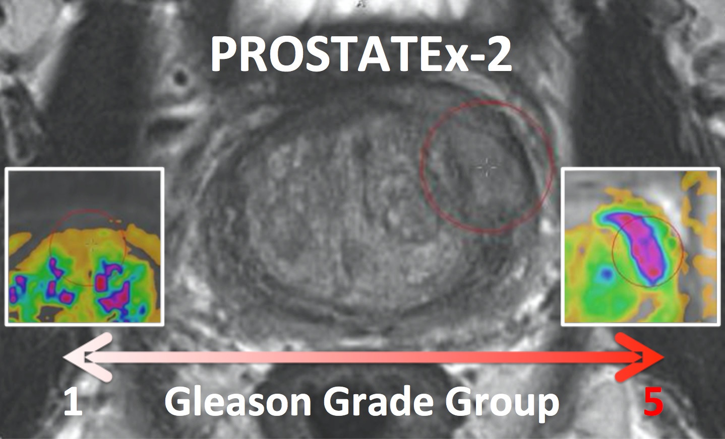 Image Added
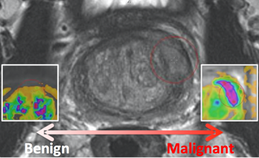 Image Added
| Excerpt |
|---|
This collection is a retrospective set of prostate MR studies. All studies included T2-weighted (T2W), proton density-weighted (PD-W), dynamic contrast enhanced (DCE), and diffusion-weighted (DW) imaging. The images were acquired on two different types of Siemens 3T MR scanners, the MAGNETOM Trio and Skyra. T2-weighted images were acquired using a turbo spin echo sequence and had a resolution of around 0.5 mm in plane and a slice thickness of 3.6 mm. The DCE time series was acquired using a 3-D turbo flash gradient echo sequence with a resolution of around 1.5 mm in-plane, a slice thickness of 4 mm and a temporal resolution of 3.5 s. The proton density weighted image was acquired prior to the DCE time series using the same sequence with different echo and repetition times and a different flip angle. Finally, the DWI series were acquired with a single-shot echo planar imaging sequence with a resolution of 2 mm in-plane and 3.6 mm slice thickness and with diffusion-encoding gradients in three directions. Three b-values were acquired (50, 400, and 800), and subsequently, the ADC map was calculated by the scanner software. All images were acquired without an endorectal coil. |
Accessing the PROSTATEx Challenge Data Sets
The PROSTATEx Challenge (" SPIE-AAPM-NCI Prostate MR Classification Challenge”) focused on quantitative image analysis methods for the diagnostic classification of clinically significant prostate
cancers and was held in conjunction with the 2017 SPIE Medical Imaging Symposium
. PROSTATEx ran from November 21, 2016 to January 15, 2017, though a "live" version has also been established at https://prostatex.grand-challenge.org which serves as an ongoing way for researchers to benchmark their performance for this task.
The PROSTATEx-2 Challenge (" SPIE-AAPM-NCI Prostate MR Gleason Grade Group Challenge" ) ran from May 15, 2017 to June 23, 2017 and was focused on the development of quantitative multi-parametric MRI biomarkers for the determination of Gleason Grade Group in prostate cancer. It was held in conjunction with the 2017 AAPM Annual Meeting (see http://www.
- Release date of training set cases with truth: November 21, 2016
- Release date of test set cases without truth: December 12, 2016
- Submission date for participants’ test set classification output: January 15, 2017
- Challenge results released to participants: January 20, 2017
- SPIE Medical Imaging Symposium: February 13-16, 2017
-2 ) .
Supplemental data and instructions specific to both challenges can be found on the Detailed Description tab below.
Acknowledgements
The prostate MR imaging was performed at the Radboud University Medical Centre (Radboudumc) in the Prostate MR Reference Center under supervision of prof. Dr. Barentsz. The Radboudumc is located in Nijmegen, The Netherlands. The dataset was collected and curated for research in computer aided diagnosis of prostate MR under supervision of Dr. Huisman, Radboudumc.
Image Removed
| Localtab Group |
|---|
| Localtab |
|---|
| active | true |
|---|
| title | Data Access |
|---|
| Data Access |
|
Note: The following download links will download the entire collection. |
|
See the Detailed Description section of this page for information about subsets specific to the PROSTATEx challenges.
| Data Type | Download all or Query/Filter | License |
|---|
Download images (15.1 GB DICOM) |
|
|
 Image Removed Image Removed Image Removed Image Removed
| Download Ktrans images (mhd format) | Image Removed |
Download lesion information (zip) | Image Removed |
| Tcia button generator |
|---|
| url | https://wiki.cancerimagingarchive.net/download/attachments/23691656/PROSTATEx-v1-doiJNLP.tcia?version=1&modificationDate=1534787029451&api=v2 |
|---|
|
|
| Tcia button generator |
|---|
| label | Search |
|---|
| url | https://www.cancerimagingarchive.net/nbia-search/?CollectionCriteria=PROSTATEx |
|---|
|
|
(Download requires NBIA Data Retriever) | | | Ktrans images (.mhd) and Lesion Info (.csv) | See Detailed Description |
| | Test set reference standard (csv, 11 kB) |
| Tcia button generator |
|---|
| url | https://wiki.cancerimagingarchive.net/download/attachments/23691656/ProstateX-Findings-Test.csv?api=v2 |
|---|
|
|
| |
|
|
Download lesion reference thumbnails (.bmp) | Image Removed
Click the Versions tab for more info about data releases. | Nci_crdc additional resources |
|---|
Third Party Analyses of this DatasetTCIA encourages the community to publish your analyses of our datasets. Below is a list of such third party analyses published using this Collection:
|
| Localtab |
|---|
| title | Detailed Description |
|---|
| Detailed Description
|
|
Clinical Data
Image Analyses (segmentations) Patients347348~4 sets of MRI scan data per case |
Prostate MR in accordance with ACR PIRADS2.0 comprises at least 3 types of images or parameters that should be jointly analyzed for the assessment of prostate cancer. The prostate MR imaging was performed at the Radboud University Medical Centre (Radboudumc) in the Prostate MR Reference Center under supervision of prof. dr. Barentsz. The Radboudumc is located in Nijmegen, The Netherlands. The dataset was collected and curated for research in computer aided diagnosis of prostate MR under supervision of dr. Huisman, Radboudumc as documented in:
G. Litjens, O. Debats, J. Barentsz, N. Karssemeijer and H. Huisman. "Computer-aided detection of prostate cancer in MRI", IEEE Transactions on Medical Imaging 2014;33:1083-1092.
If you use this data for research than refer the above publication.
PROSTATEx Challenge (November 21, 2016 to February 16, 2017)SPIE, along with the support of the American Association of Physicists in Medicine (AAPM) and the National Cancer Institute (NCI), conducted a “Grand Challenge” on quantitative image analysis methods for the diagnostic classification of clinically significant prostate lesions. For more details, go to https://prostatex.grand-challenge.org/ . Training and test cohorts along with supplemental Ktrans images and lesion information can be obtained separately using the following options for download:
| Data Type | Training Cohort (204 subjects) | Test Cohort (140 subjects) |
|---|
Download images (DICOM, 126 kB) (DICOM, 121 kB)
|
| Tcia button generator |
|---|
| url | https://wiki.cancerimagingarchive.net/download/attachments/23691656/PROSTATEx-train.tcia?version=1&modificationDate=1534787030035&api=v2 |
|---|
|
|
|
| Tcia button generator |
|---|
| url | https://wiki.cancerimagingarchive.net/download/attachments/23691656/PROSTATEx-test.tcia?version=1&modificationDate=1534787450806&api=v2 |
|---|
|
|
| Download Ktrans images (.mhd 161 kB) (.mhd 119 kB) |
| Tcia button generator |
|---|
| url | https://app.box.com/s/y871i386j4o9rqwcsms5mqni63nojzx1 |
|---|
|
|
|
| Tcia button generator |
|---|
| url | https://app.box.com/s/k3iofc0r3ktjnb4f7105lssdzjon95ie |
|---|
|
|
| Download lesion information (.zip, 281 kB) (zip, 236 kB) |
| Tcia button generator |
|---|
| url | https://wiki.cancerimagingarchive.net/download/attachments/23691656/ProstateX-TrainingLesionInformationv2.zip?version=2&modificationDate=1483479231532&api=v2 |
|---|
|
|
|
| Tcia button generator |
|---|
| url | https://wiki.cancerimagingarchive.net/download/attachments/23691656/ProstateX-TestLesionInformation.zip?version=2&modificationDate=1483479234096&api=v2 |
|---|
|
|
| Download lesion reference thumbnails (.bmp, 37.6 MB) (.bmp, 23.3 MB) |
| Tcia button generator |
|---|
| url | https://wiki.cancerimagingarchive.net/download/attachments/23691656/ProstateX-Screenshots-Train.zip?version=1&modificationDate=1479401241653&api=v2 |
|---|
|
|
|
| Tcia button generator |
|---|
| url | https://app.box.com/s/7jn4jtd3pbi9rlurc4pvnkx554lnfljw |
|---|
|
|
|
The images come in two encodings. The acquired MR is provided in DICOM encoding. Additionally Ktrans images are provided. They come in mhd format. Ktrans is a key pharmacokinetic parameter computed from the available Dynamic contrast enhanced T1-weighted series. Each patient has one study with several DICOM images and one Ktrans image. The Ktrans image is encoded in two files ProstateX-[ProxID]-Ktrans.[mhd/zraw], where ProxID is the ProstateX patient identifier. The DICOM images comprise several Series each comprising several Instances. The DICOM files are documented in the ProstateX-Images.csv file. The columns in that file encode the following: ProxID – ProstateX patient identifier. Name – Series Description Studydate – Study Date fid – Finding ID Pos – Scanner Coordinate position of the finding WorldMatrix – Matrix describing image orientation and scaling ijk – image col,row,slice coordinate of finding ImageUID – Image Identifier TopLevel 0 - Series forms one image 1 – A set of Series forms a 4D image (e.g. Dynamic MR) NA – Series form one image, but is part of a Level 1 4D image
SpacingBetweenSlices – Scalar Spacing between slices VoxelSpacing – Vector with x,y,z spacing scalars Dim – Vector with 4D dimensions of the image DCMSerDescr – The original DICOM Series Description DCMSerUID – The DICOM Series UID DCMSerNum – The DICOM Series Number InstanceUIDList – DICOM Instances that make up this series ImageUIDList – TopLevel-NA Images the make up this Toplevel 1 image
For example, to get the ADC image of Patient ProstateX-0123 do the following. After you imported the DICOM files into your environment, |
|
go to patient ProstateX-0123 and find the series with ADC in it. In this case it is ‘ep2d_diff_tra_DYNDIST_ADC’. It has SeriesNumber 8. The DICOM images in that series form the ADC image for this challenge. Image slice j at coordinate i,j contains a finding fid. See findings for more details. Findings The findings are documented in the ProstateX-Findings.csv table. Documentation for the columns in that table is as follows: pos - Scanner Coordinate position of the finding ClinSig – Identifier available in training set that identifies whether this is a clinically significant finding. Either the biopsy GleasonScore was 7 or higher. Findings with a PIRADS score 2 were not biopsied and are not considered clinically significant. In our center the occurrence of clinically significant cancer in PIRADS 2 lesions is less than 5%.
|
|
| Localtab |
|---|
| title | Citations & Data Usage Policy |
|---|
|
Citations & Data Usage Policy
This collection is freely available to browse, download, and use for commercial, scientific and educational purposes as outlined in the Creative Commons Attribution 3.0 Unported License. See TCIA's Data Usage Policies and Restrictions for additional details. Questions may be directed to help@cancerimagingarchive.net.
Please be sure to include the following citations in your work if you use this data set:Note, ProxID is the PROSTATEx case ID, and fid is the finding (i.e., lesion) ID in both the ProstateX-Findings.csv file and the ProstateX-Images.csv file. The Findings spreadsheet has one row per lesion (if a case has only one lesion, then the only fid for that case will be “1” , alternately if a case has two lesions, then there will be an fid of “1” and an fid of “2” for that case). The Images spreadsheet has a row for every image that contains the lesion - that is why there are multiple rows with the same (ProxID, fid) combination.
PROSTATEx-2 — SPIE-AAPM-NCI Prostate MR Gleason Grade Group Challenge (May 15, 2017 to August 3, 2017)The American Association of Physicists in Medicine (AAPM), along with the SPIE (the international society for optics and photonics) and the National Cancer Institute (NCI), conducted a part 2 “Grand Challenge” on the development of quantitative multi-parametric magnetic resonance imaging (MRI) biomarkers for the determination of Gleason Grade Group in prostate cancer. For more details about PROSTATEx-2 please go to http://www.aapm.org/GrandChallenge/PROSTATEx-2/default.asp . Training and test cohorts along with supplemental Ktrans images and lesion information can be obtained separately using the following options for download:
| Data Type | Training Cohort (112 subjects) | Test Cohort (70 subjects) |
|---|
Download images (DICOM) |
| Tcia button generator |
|---|
| url | https://wiki.cancerimagingarchive.net/download/attachments/23691656/PROSTATExChallenge2-v2-doiJNLP.tcia?version=1&modificationDate=1534787028529&api=v2 |
|---|
|
|
|
| Tcia button generator |
|---|
| url | https://wiki.cancerimagingarchive.net/download/attachments/23691656/PROSTATEx2-test.tcia?version=1&modificationDate=1534787027933&api=v2 |
|---|
|
|
| Download Ktrans images (.mhd) |
| Tcia button generator |
|---|
| url | https://app.box.com/s/xv6j8yhq4nd7c1eqdn88n1maw3un0xbv |
|---|
|
|
|
| Tcia button generator |
|---|
| url | https://app.box.com/s/ibue53ch74tst8hd7hknvhw01jxruw2v |
|---|
|
|
| Download lesion information (.zip) |
| Tcia button generator |
|---|
| url | https://wiki.cancerimagingarchive.net/download/attachments/23691656/ProstateX2-DataInfo-Train.zip?version=1&modificationDate=1494608672116&api=v2 |
|---|
|
|
|
| Tcia button generator |
|---|
| url | https://wiki.cancerimagingarchive.net/download/attachments/23691656/ProstateX2-DataInfo-Test.zip?version=1&modificationDate=1496674918589&api=v2 |
|---|
|
|
| Download lesion reference thumbnails (.bmp) |
| Tcia button generator |
|---|
| url | https://wiki.cancerimagingarchive.net/download/attachments/23691656/ProstateXChallenge2ScreenshotsTrain.zip?version=1&modificationDate=1494518725752&api=v2 |
|---|
|
|
|
| Tcia button generator |
|---|
| url | https://wiki.cancerimagingarchive.net/download/attachments/23691656/ProstateXChallenge2ScreenshotsTest.zip?version=1&modificationDate=1496674921348&api=v2 |
|---|
|
|
|
The training set will consist of 112 findings. This dataset will be representative of the technical properties (scanner type, acquisition parameters, file format) and the nature of the prostate lesions of the test set. An associated Excel file will include case name, the coordinates of the centroid of all lesions, and the lesion ‘truth’ label (Gleason Grade Group). Reference thumbnail images of the lesions will also be provided. The test set will consist of 70 findings. The locations of the lesions will be specified in the accompanying Excel file that will follow the same format as for the training set with the omission of the ‘truth’ labels. Reference thumbnail images of the lesions will also be provided.
|
| Localtab |
|---|
| title | Citations & Data Usage Policy |
|---|
| Citations & Data Usage Policy | Public collection license |
|---|
|
|
| Info |
|---|
| title | Publication Citation |
|---|
| Litjens, G. |
|
|
Aided Detection of Prostate Cancer in MRI. In IEEE Transactions on Medical |
|
|
| Info |
|---|
| Clark K, Vendt B, Smith K, Freymann J, Kirby J, Koppel P, Moore S, Phillips S, Maffitt D, Pringle M, Tarbox L, Prior F. The Cancer Imaging Archive (TCIA): Maintaining and Operating a Public Information Repository, Journal of Digital Imaging, Volume 26, Number 6, December, 2013, pp 1045-1057 |
|
|
data. If you have a publication you'd like to add, please contact |
|
TCIA's Helpdesk. | Html-bobswift |
|---|
<iframe class="airtable-embed" src="https://airtable.com/embed/shrIUQYW4LQU8WvFu?backgroundColor=purple&layout=card" frameborder="0" onmousewheel="" width="100%" height="533" style="background: transparent; border: 1px solid #ccc;"></iframe> |
|
|
05
| Data Type | Download all or Query/Filter | License |
|---|
Download images (15.1 GB DICOM) |
|
|
Image Removed Image Removed
| Download Ktrans images (mhd format) | Image Removed |
Download lesion information (zip) | Image Removed |
Download lesion reference thumbnails (.bmp) | Image Removed |
|
| Tcia button generator |
|---|
| url | https://wiki.cancerimagingarchive.net/download/attachments/23691656/PROSTATEx-v1-doiJNLP.tcia?version=1&modificationDate=1534787029451&api=v2 |
|---|
|
|
| Tcia button generator |
|---|
| label | Search |
|---|
| url | https://nbia.cancerimagingarchive.net/nbia-search/?CollectionCriteria=PROSTATEx |
|---|
|
|
(Download requires NBIA Data Retriever) | | | Ktrans images (.mhd) and Lesion Info (.csv) | See Detailed Description |
| | Test set reference standard (csv) |
| Tcia button generator |
|---|
| url | https://wiki.cancerimagingarchive.net/download/attachments/23691656/ProstateX-Findings-Test.csv?api=v2 |
|---|
|
|
| |
Added Test Reference Standard document Version 1: Updated 2017/03/29
| Data Type | Download all or Query/Filter |
|---|
Download images (15.1 GB DICOM) |
| Tcia button generator |
|---|
| url | https://wiki.cancerimagingarchive.net/download/attachments/23691656/PROSTATEx-v1-doiJNLP.jnlp?version=1&modificationDate=1490928917140&api=v2 |
|---|
|
|
| Tcia button generator |
|---|
| label | Search |
|---|
| url | https://nbia.cancerimagingarchive.net/ncia/externalLinks.jsf?collectionName=PROSTATEx |
|---|
|
|
|
|
|