Versions Compared
Key
- This line was added.
- This line was removed.
- Formatting was changed.
Summary
| Excerpt |
|---|
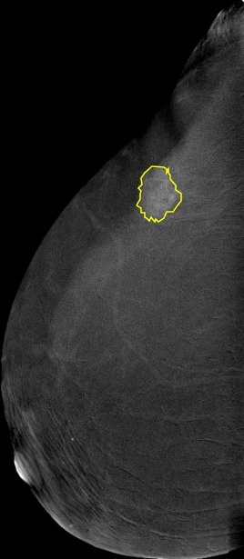
Contrast-enhanced spectral mammography (CESM) is done using the standard digital mammography equipment, with additional software that performs dual-energy image acquisition. The dataset is a collection of 2006 CESM images all high resolution with an average of 2355 x 1315 pixels. Each image with its corresponding manual annotation (breast composition, mass shape, mass margin, mass density, architectural distortion, asymmetries, calcification type, calcification distribution, mass enhancement pattern, non-mass enhancement pattern, non-mass enhancement distribution, and overall BIRADS assessment) is compiled into 1 Excel file. Moreover, full medical reports are provided for each case (DOCX) along with manual segmentation annotation for the abnormal findings in each image (CSV file). Deep learning (DL) has a promising potential in reducing the workload of radiologists and helping them provide a more accurate diagnosis. However, fully annotated and large-sized datasets are required. In the past couple of years, public mammography datasets were released. These datasets contain digital mammography images only, and none include CESM images. Acquisition protocol: Two minutes after intravenously injecting the patient with non-ionic low-osmolar iodinated contrast material (dose: 1.5 mL/kg), craniocaudal (CC) and mediolateral oblique (MLO) views are obtained. Each view comprises two exposures, one with low energy (peak kilo-voltage values ranging from 26 to 31kVp) and one with high energy (45 to 49 kVp). Low and high-energy images are then recombined and subtracted through appropriate image processing to suppress the background breast parenchyma. A complete examination is carried out in about 5-6 minutes. Image preprocessing: The images were converted from DICOM to JPEG using RadiAnt with best 100% image quality (lossless). |
Acknowledgements
We would like to acknowledge the individuals and institutions that have provided data for this collection:
- National Cancer Institute, Cairo University, Cairo, Egypt : Special thanks to Dr. Rana Khaled, M.Sc, Prof. Maha Helal, MD, Prof. Omnia Mokhtar, MD and Dr. Hebatalla El Kassas, MD from the Department of Radiology.
- Faculty of Computers and Artificial Intelligence, Cairo University, Cairo, Egypt – Special thanks to Omar Alfarghaly, Prof. Abeer Elkorany, and Prof. Aly Fahmy from the Department of Computer Science.
| Localtab Group | ||||||||||||||||||||||||||||||||||||||||||||||||||||||||||||||||||||||||||||||||||||||||||||||||||||||
|---|---|---|---|---|---|---|---|---|---|---|---|---|---|---|---|---|---|---|---|---|---|---|---|---|---|---|---|---|---|---|---|---|---|---|---|---|---|---|---|---|---|---|---|---|---|---|---|---|---|---|---|---|---|---|---|---|---|---|---|---|---|---|---|---|---|---|---|---|---|---|---|---|---|---|---|---|---|---|---|---|---|---|---|---|---|---|---|---|---|---|---|---|---|---|---|---|---|---|---|---|---|---|
|
