| Localtab |
|---|
| active | true |
|---|
| title | Data Access |
|---|
| Data AccessClick the Download button to save a ".tcia" manifest file to your computer, which you must open with the NBIA Data Retriever. Click the Search button to open our Data Portal, where you can browse the data collection and/or download a subset of its contents.| Data Type | Download all or Query/Filter | License |
|---|
| Images, Segmentations, and Radiation Therapy Structures (DICOM, |
29GB) | Lung1 clinical (CSV) | | 33 GB) |
| Tcia button generator |
|---|
| url | https://wiki.cancerimagingarchive.net/download/attachments/16056854/NSCLC%20Radiomics%20Version%204%20Oct%202020%20NBIA-manifest.tcia?version=1&modificationDate=1603198863613&api=v2 |
|---|
|
|
| Tcia button generator |
|---|
| label | Search |
|---|
| url | https://nbia.cancerimagingarchive.net/nbia-search/?MinNumberOfStudiesCriteria=1&CollectionCriteria=NSCLC-Radiomics |
|---|
|
|
(Download requires the NBIA Data Retriever) | | | Lung1 clinical (CSV, 23 kB) |
| Tcia button generator |
|---|
| url | https://wiki.cancerimagingarchive.net/download/attachments/16056854/NSCLC%20Radiomics%20Lung1.clinical-version3-Oct%202019.csv?version=1&modificationDate=1572013183040&api=v2 |
|---|
|
|
| |
 Image Removed Image RemovedClick the Versions tab for more info about data releases. | Nci_crdc additional resources |
|---|
Third Party Analyses of this DatasetTCIA encourages the community to publish your analyses of our datasets. Below is a list of such third party analyses published using this Collection: |
| Localtab |
|---|
| title | Detailed Description |
|---|
| Detailed Description | |
|---|
Modalities | CT, RTSTRUCT, SEG | Number of PatientsParticipants | 422 | Number of Studies | 844422 | Number of Series | 1265 | Number of Images | 5207252073 | | Image Size (GB) | 29.333 |
Radiation Oncologist Tumor Segmentations
The DICOM Radiotherapy Structure Sets (RTSTRUCT) and DICOM Segmentation (SEG) files in this data contain a manual delineation by a radiation oncologist of the 3D volume of the primary gross tumor volume ("GTV-1"). For viewing quickly we recommend Dicompyler (http://www.dicompyler.com/) which is ) and selected anatomical structures (i.e., lung, heart and esophagus). Of note, DICOM SEG objects contain a subset of annotations available in RTSTRUCT.
For viewing the annotations the authors recommend 3D Slicer that can be used to view both RTSTRUCT and SEG annotations (make sure you install the SlicerRT and QuantitativeReporting extensions first!). Visualization of the DICOM annotations is also supported by the OHIF Viewer.
Other tools include: - Dicompyler is an open source, cross-platform DICOM RT viewer.
Slicer has a SlicerRT module (http://slicerrt.github.io/index.html) which enables use of this kind of data. The (https://github.com/dicom/rtkit- .
- dcmqi and Plastimatch libraries can be used to support conversion of the DICOM SEG and RTSTRUCT representations, respectively, into the popular research formats, such as NIfTI or NRRD
)Clinical DataCorresponding clinical data can be found here: Lung1.clinical.csv. Please note that survival time is measured in days from start of treatment. DICOM patients names are identical in TCIA and clinical data file. The deadstatus.event follows the standard epidemiological / biomedical definition for time to event survival modelling whereby : 1 == “death has occurred at the time interval value stated on the next column”, hence 0 == “death has NOT occurred up until the time interval value stated in the next column, and is therefore right-censored”. Note:Some CT ( LUNG1-014 , LUNG1-021 , LUNG1-085) are missing some slices from the complete volume, unfortunately not detected until recently. The skipped CT slice did not cut through the Gross Tumour Volume which was the principal focus of this Collection. We understand the user may have a different principal focus for re-using this collection, but perhaps some other workaround like interpolation may be timely and feasible for those affected volumes. |
| Localtab |
|---|
| title | Citations & Data Usage Policy |
|---|
| Citations & Data Usage Policy This collection may not be used for commercial purposes. It is available to browse, download, as outlined in the Attribution-NonCommercial 3.0 Unported (CC BY-NC 3.0) https://creativecommons.org/licenses/by-nc/3.0/. See TCIA's Data Usage Policies and Restrictions for additional details. Questions may be directed to help@cancerimagingarchive.net. Please be sure to include the following citations in your work if you use this data set: | Tcia limited license policy |
|---|
| Info |
|---|
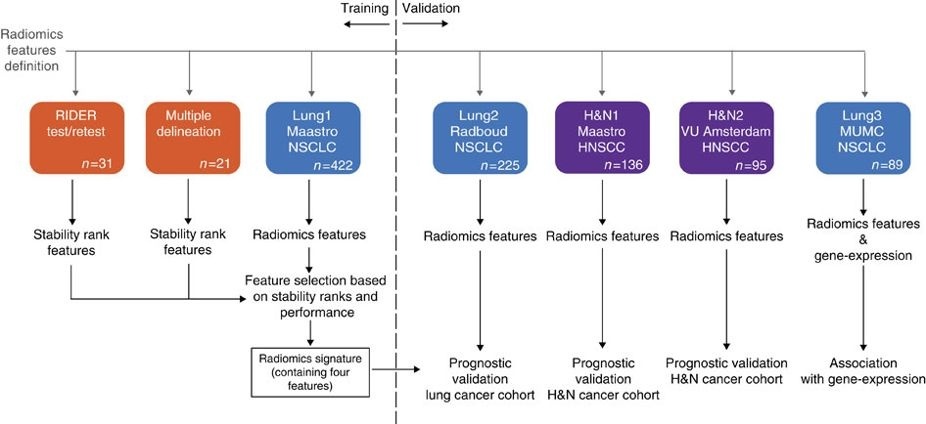
| Aerts, H. J. W. L., Wee, L., Rios Velazquez, E., Leijenaar, R. T. H., Parmar, C., Grossmann, P., Carvalho, S., Bussink, J., Monshouwer, R., Haibe-Kains, B., Rietveld, D., Hoebers, F., Rietbergen, M. M., Leemans, C. R., … , Dekker, A., Quackenbush, J., Gillies, R. J., Lambin, P. (20192014). Data From NSCLC-Radiomics (version 4) [Data set]. The Cancer Imaging Archive. https://doi.org/10.7937/K9/TCIA.2015.PF0M9REI |
| Info |
|---|
| title | Publication Citation |
|---|
| Aerts, H. J. W. L., Velazquez, E. R., Leijenaar, R. T. H., Parmar, C., Grossmann, P., CavalhoCarvalho, S., … Bussink, J., Monshouwer, R., Haibe-Kains, B., Rietveld, D., Hoebers, F., Rietbergen, M. M., Leemans, C. R., Dekker, A., Quackenbush, J., Gillies, R. J., Lambin, P. (2014, June 3). Decoding tumour phenotype by noninvasive imaging using a quantitative radiomics approach. Nature Communications. Nature Publishing Group.http https://doi.org/10.1038/ncomms5006 (link) |
| Info |
|---|
| Clark K, Vendt B, Smith K, Freymann J, Kirby J, Koppel P, Moore S, Phillips S, Maffitt D, Pringle M, Tarbox L, Prior F. The Cancer Imaging Archive (TCIA): Maintaining and Operating a Public Information Repository, Journal of Digital Imaging, Volume 26, Number 6, December, 2013, pp 1045-1057. (paper). https://doi.org/10.1007/s10278-013-9622-7 |
Questions may be directed to help@cancerimagingarchive.net. Other Publications Using This DataTCIA maintains a list of publications that which leverage our data. If you have a publication you'd like to add, please contact the TCIA Helpdesk. |
| Localtab |
|---|
| Version 3 4 (Current): Updated 2020/10/22| Data Type | Download all or Query/Filter |
|---|
| Images (DICOM, 33 GB) |
| Tcia button generator |
|---|
| url | https://wiki.cancerimagingarchive.net/download/attachments/16056854/NSCLC%20Radiomics%20Version%204%20Oct%202020%20NBIA-manifest.tcia?version=1&modificationDate=1603198863613&api=v2 |
|---|
|
|
| Tcia button generator |
|---|
| label | Search |
|---|
| url | https://nbia.cancerimagingarchive.net/nbia-search/?MinNumberOfStudiesCriteria=1&CollectionCriteria=NSCLC-Radiomics |
|---|
|
|
(Download requires the NBIA Data Retriever) | | Lung1 clinical (CSV) |
| Tcia button generator |
|---|
| url | https://wiki.cancerimagingarchive.net/download/attachments/16056854/NSCLC%20Radiomics%20Lung1.clinical-version3-Oct%202019.csv?version=1&modificationDate=1572013183040&api=v2 |
|---|
|
|
|
- RTSTRUCT and SEG study instance UID changed to match study instance uid with associated CT image.
- Added missing structures in SEG files to match associated RTSTRUCTs.
- Patient Id copied to Patient Name in CT images (for consistency).
- Added 1 missing image for LUNG1-246.
Version 3: Updated 2019/10/23| Data Type | Download all or Query/Filter |
|---|
| Images (DICOM, 29GB) |
| Tcia button generator |
|---|
| url | https://wiki.cancerimagingarchive.net/download/attachments/16056854/NSCLC-Radiomics-V3-Oct2019.NBIA-manifest.tcia?version=1&modificationDate=1571863634068&api=v2 |
|---|
|
|
(Download requires the NBIA Data Retriever Image Removed  Image Removed Image Removed (Requires the NBIA Data Retriever.) | | Lung1 clinical (CSV) |
| Tcia button generator |
|---|
| url | https://wiki.cancerimagingarchive.net/download/attachments/16056854/NSCLC%20Radiomics%20Lung1.clinical-version3-Oct%202019.csv?version=1&modificationDate=1572013183040&api=v2 |
|---|
|
Image Removed
|
- Re-checked and updated the RTSTRUCT files to amend issues in the previous submission due to missing RTSTRUCTS or regions of interest that were not vertically aligned with the patient image.
- In 4 cases (LUNG1-083,LUNG1-095,LUNG1-137,LUNG1-246) re-submitted the correct CT images.
- The regions of interest now include the primary lung tumor labelled as “GTV-1”, as well as organs at risk.
- For one case (LUNG1-128) the subject does not have GTV-1 because it was actually a post-operative case; we retained the CT scan here for completeness.
- Added DICOM SEGMENTATION objects to the collection, which makes it easier to search and retrieve the GTV-1 binary mask for re-use in quantitative imaging research.
- Clinical data updated as follow-up time has been extended.
Version 2: Updated 2016/05/3131 | Data Type | Download all or Query/Filter |
|---|
| Images (DICOM, 25GB) |  Image Removed Image Removed Image Removed Image Removed
(Requires the NBIA Data Retriever.) not available, see version 3 | | Lung1 clinical (CSV) |
| Tcia button generator |
|---|
| url | https://wiki.cancerimagingarchive.net/download/attachments/16056854/Lung1.clinical_%20version%201%20and%20version2.csv?version=1&modificationDate=1584901749543&api=v2 |
|---|
|
Image Removed
|
Added 318 RTSTRUCT files for existing subject imaging data. (NOTE: version 2 removed as RTSTRUCTs or regions of interest were not vertically aligned with patient images. See version 3 for updated files). Version 1: Updated 2014/07/02| Data Type | Download all or Query/Filter |
|---|
| Images (DICOM, 25GB) |
| Tcia button generator |
|---|
| url | https://wiki.cancerimagingarchive.net/download/attachments/16056854/TCIA_NSCLC-Radiomics_06-22-2015.tcia?version=1&modificationDate=1534787429158&api=v2 |
|---|
|
|
(Download requires the NBIA Data Retriever) Image Removed Image Removed the NBIA Data Retriever. | | Lung1 clinical (CSV) |
| Tcia button generator |
|---|
| url | https://wiki.cancerimagingarchive.net/download/attachments/16056854/Lung1.clinical_%20version%201%20and%20version2.csv?version=1&modificationDate=1584901749543&api=v2 |
|---|
|
Image Removed
|
|
|