Child pages
  • SPIE-AAPM-NCI PROSTATEx Challenges (PROSTATEx)

Versions Compared

Key

  • This line was added.
  • This line was removed.
  • Formatting was changed.

Summary


Excerpt

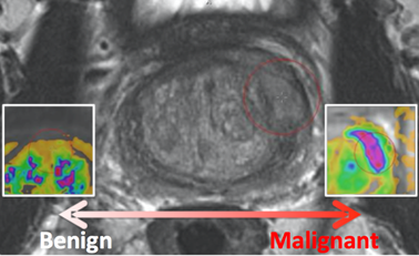
SPIE, along with the support of the American Association of Physicists in Medicine (AAPM) and the National Cancer Institute (NCI), will conduct a “Grand Challenge” on quantitative image analysis methods for the diagnostic classification of clinically significant prostate lesions.  As part of the 2017 SPIE Medical Imaging Symposium, the PROSTATEx Challenge will provide a unique opportunity for participants to compare their algorithms with those of others from academia, industry, and government in a structured, direct way using the same data sets.  For more details, go to http://www.spie.org/PROSTATEx/.  To register for the challenge visit http://spiechallenges.cloudapp.net/

  • Release date of training set cases with truth:  November 21, 2016
  • Release date of test set cases without truth:  December 12, 2016
  • Submission date for participants’ test set classification output:  January 16, 2017
  • Challenge results released to participants:  January 20, 2017
  • SPIE Medical Imaging Symposium:  February 13-16, 2017

Localtab Group
Localtab
activetrue
titleData Access

Data Access

Choosing the Download option will provide you with a file to launch the TCIA Download Manager to download the entire collection. If you want to browse or filter the data to select only specific scans/studies please use the Search By Collection option.

Data Type
Download all or Query/Filter

Images (DICOM, 15.1GB)

Excel workbook file with clinical and outcome data for all subjects 


The PROSTATEx Challenge Organizers have provided the following options for download. The significance of these subsets are explained on the the challenge web site: http://spiechallenges.cloudapp.net/competitions/6.  You may also search/filter the data via our Data Portal if you are only looking for a subset of this data.

Data Type

Training Cohort

(204 subjects)

Test Cohort

(140 subjects)

Download images (DICOM)


Download Ktrans images (.mhd)

KTrans-Training-Files  KTrans-Training-Files

Download lesion information (.zip)


Download lesion reference thumbnails (.bmp)

Click the Versions tab for more info about data releases.

Localtab
titleDetailed Description

Detailed Description

Collection Statistics


Modalities

MR

Number of Patients

346

Number of Studies

349

Number of Series

18,321

Number of Images

309,251

Images Size (GB)15.1


Prostate MR in accordance with ACR PIRADS2.0 comprises at least 3 types of images or parameters that should be jointly analyzed for the assessment of prostate cancer. The prostate MR imaging was performed at the Radboud University Medical Centre (Radboudumc) in the Prostate MR Reference Center under supervision of prof. dr. Barentsz. The Radboudumc is located in Nijmegen, The Netherlands. The dataset was collected and curated for research in computer aided diagnosis of prostate MR under supervision of dr. Huisman, Radboudumc as documented in:

Info
titleCitation
G. Litjens, O. Debats, J. Barentsz, N. Karssemeijer and H. Huisman. "Computer-aided detection of prostate cancer in MRI", IEEE Transactions on Medical Imaging 2014;33:1083-1092.

If you use this data for research then refer the above publication.

The images come in two encodings. The acquired MR is provided in DICOM encoding. Additionally Ktrans images are provided. They come in mhd format. Ktrans is a key pharmacokinetic parameter computed from the available Dynamic contrast enhanced T1-weighted series. Each patient has one study with several DICOM images and one Ktrans image. The Ktrans image is encoded in two files ProstateX-[ProxID]-Ktrans.[mhd/zraw], where ProxID is the ProstateX patient identifier. The DICOM images comprise several Series each comprising several Instances. The DICOM files are documented in the ProstateX-Images.csv file. The columns in that file encode the following:

  • ProxID – ProstateX patient identifier.

  • Name – Series Description

  • Studydate – Study Date

  • fid – Finding ID

  • Pos – Scanner Coordinate position of the finding

  • WorldMatrix – Matrix describing image orientation and scaling

  • ijk – image col,row,slice coordinate of finding

  • ImageUID – Image Identifier

  • TopLevel

    • 0 - Series forms one image

    • 1 – A set of Series forms a 4D image (e.g. Dynamic MR)

    • NA – Series form one image, but is part of a Level 1 4D image

  • SpacingBetweenSlices – Scalar Spacing between slices

  • VoxelSpacing – Vector with x,y,z spacing scalars

  • Dim – Vector with 4D dimensions of the image

  • DCMSerDescr – The original DICOM Series Description

  • DCMSerUID – The DICOM Series UID

  • DCMSerNum – The DICOM Series Number

  • InstanceUIDList – DICOM Instances that make up this series

  • ImageUIDList – TopLevel-NA Images the make up this Toplevel 1 image

For example, to get the ADC image of Patient ProstateX-0123 do the following. After you imported the DICOM files into your environment, go to patient ProstateX-0123 and find the series with ADC in it. In this case it is ‘ep2d_diff_tra_DYNDIST_ADC’. It has SeriesNumber 8. The DICOM images in that series form the ADC image for this challenge. Image slice j at coordinate i,j contains a finding fid. See findings for more details.

Findings

The findings are documented in the ProstateX-Findings.csv table. Documentation for the columns in that table is as follows:

  • ProxID – ProstateX patient identifier

  • fid - Finding ID

  • pos - Scanner Coordinate position of the finding

  • ClinSig – Identifier available in training set that identifies whether this is a clinically significant finding. Either the biopsy GleasonScore was 7 or higher. Findings with a PIRADS score 2 were not biopsied and are not considered clinically significant. In our center the occurrence of clinically significant cancer in PIRADS 2 lesions is less than 5%.

Localtab
titleCitations & Data Usage Policy

Citations & Data Usage Policy 

This collection is freely available to browse, download, and use for commercial, scientific and educational purposes as outlined in the Creative Commons Attribution 3.0 Unported License.  See TCIA's Data Usage Policies and Restrictions for additional details. Questions may be directed to help@cancerimagingarchive.net.

Please be sure to include the following citations in your work if you use this data set:

Info
titleData Citation

Geert Litjens, Oscar Debats, Jelle Barentsz, Nico Karssemeijer, and Henkjan Huisman. ProstateX Challenge data. <doi coming soon> https://doi.org/10.7937/K9TCIA.2017.MURS5CL

Info
titlePublication Citation

G. Litjens, O. Debats, J. Barentsz, N. Karssemeijer and H. Huisman. "Computer-aided detection of prostate cancer in MRI", IEEE Transactions on Medical Imaging 2014;33:1083-1092.

Info
titleTCIA Citation

Clark K, Vendt B, Smith K, Freymann J, Kirby J, Koppel P, Moore S, Phillips S, Maffitt D, Pringle M, Tarbox L, Prior F. The Cancer Imaging Archive (TCIA): Maintaining and Operating a Public Information Repository, Journal of Digital Imaging, Volume 26, Number 6, December, 2013, pp 1045-1057. (paper)

Other Publications Using This Data

TCIA maintains a list of publications which leverage our data. At this time we are not aware of any publications based on this data. If you have a publication you'd like to add please contact the TCIA Helpdesk.

Localtab
titleVersions

Version 1 (Current): Updated 2017/03/29

Data TypeDownload all or Query/Filter

Download images (15.1 GB DICOM)


Download Ktrans images (mhd format)


Download lesion information (zip)


Download lesion reference thumbnails (.bmp)