Summary
| Redirect | ||||
|---|---|---|---|---|
|
| Excerpt |
|---|
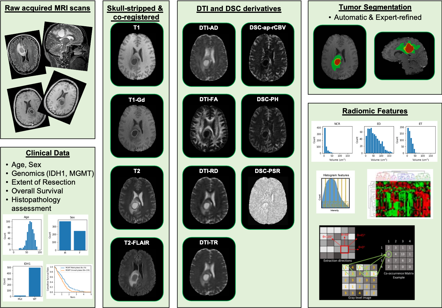
This collection comprises multi-parametric magnetic resonance imaging (mpMRI) scans for de novo Glioblastoma (GBM) patients from the University of Pennsylvania Health System, coupled with patient demographics, clinical outcome (e.g., overall survival, genomic information, tumor progression), as well as computer-aided and manually-corrected segmentation labels of multiple histologically distinct tumor sub-regions, computer-aided and manually-corrected segmentations of the whole brain, a rich panel of radiomic features along with their corresponding co-registered mpMRI volumes in NIfTI format. Scans were initially skull-stripped and co-registered, before their tumor segmentation labels were produced by an automated computational method. These segmentation labels were revised and any label misclassifications were manually corrected/approved by expert board-certified neuroradiologists. The final labels were used to extract a rich panel of imaging features, including intensity, volumetric, morphologic, histogram-based and textural parameters. The segmentation labels enable quantitative computational and clinical studies without the need to repeat manual annotations whilst allowing for comparison across studies. They can also serve as a set of manually-annotated gold standard labels for performance evaluation in computational challenges. The provided panel of radiomic features may facilitate research integrative of the molecular characterization offered, and hence allow associations with molecular markers (radiogenomic biomarker research), clinical outcomes, treatment responses and other endpoints, by researchers without sufficient computational background to extract such features. Additional data accompanying the UPENN-GBM data collection include H&E-stained digitized tissue sections from resected tumor specimens of matched de novo and recurrent cases for a few of the patients in this collection. |
...