Summary
In 2004 when presenting the NCI Executive Committee the ACRIN proposal to conduct the CTC trial, a case was made that publicly accessible image data sharing would offer a valuable research asset to a wide image processing research community. Adding to the many merits of that proposal, the data-sharing component was strongly endorsed. ACRIN completed the trial expeditiously and its results were published in NEJM in fall 2008 to wide interest. ACRIN has graciously allowed the wider research community access to a portion of the data from that trial here on TCIA, including spreadsheets identifying positive and negative polyp cases. For more information about these images please see the following:
- NEJM publication for their published results (N Engl J Med. 2008 Sep 18;359(12):1207-17)
- Complete ACRIN protocol description (http://www.acrin.org/TabID/151/Default.aspx).
Data Access
Imaging Data
| Info |
|---|
You can view and download these images on The Cancer Imaging Archive by logging in to TCIA and selecting the CT Colonography collection. |
Collection Statistics |
|
|---|---|
Modalities | CT |
Number of Patients | 825 |
Number of Studies | 836 |
Number of Series | 3,451 |
Number of Images | 941,774 |
If you are unsure how to download this Collection please view our quick guide on Searching by Collection or refer to our The Cancer Imaging Archive User's Guide for more detailed instructions on using the site.
Metadata
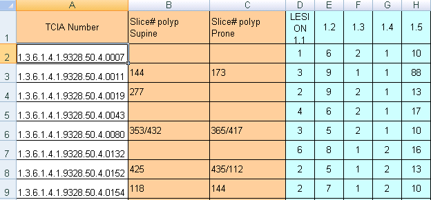
There are presently 825 cases in this collection with XLS sheets that provide polyp descriptions and their location within the colon segments. To link the XLS polyp tables with the DICOM image studies in TCIA you should understand that some cases in the TCIA are identified by long numbers with the last 4 digits after the last decimal point (e.g.: NCIA study number "1.3.6.1.4.1.9328.50.4.0040" referred to as case "40"). In addition there are a fewer number of additional positive cases that begin their identification number with 'CTC' (e.g.: CTC-5401799343)
...
You will note that two XLS files with positive findings have multiple columns descriptors of individual polyp lesions listed as in the table below. The meaning of the colored columns labeled "LESION 1.1...1.2...1.3...1.4, etc" is explained in the attached key-code ".tiff" file entitled "Polyp description key table.tiff"). Some CT scan slice numbers where the polyps were found are provided, but unfortunately the table may not have complete slice number information – you'll just have to do the best you can with the data NCI was given.
Data Access
Collection Statistics |
|
|---|---|
Modalities | CT |
Number of Patients | 825 |
Number of Studies | 836 |
Number of Series | 3,451 |
Number of Images | 941,774 |
You can view and download these images on the Cancer Imaging Archive by selecting the CT Colonography collection. If you are unsure how to download this Collection view our quick guide on Searching by Collection or you can refer to our The Cancer Imaging Archive User's Guide for more detailed instructions on using the site.