Summary
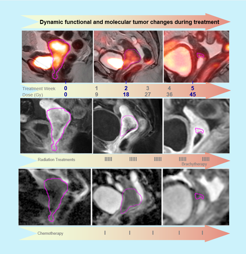
Functional/molecular imaging offers the opportunity to assess biological heterogeneity across the entire tumor volume (spatially) and longitudinally across the treatment course (temporally). In advanced cervical cancer, the tumor gradually undergoes radiation/chemotherapy induced functional and biological changes within its heterogeneous volume that can be assessed by sequential imaging before and during treatment.
Advanced cervical cancer is an ideal disease to study – in clinical patients – the vascular, cellular and molecular tumor properties that can provide essential information to monitor therapeutic responsiveness, facilitate treatment planning and may provide early prediction of ultimate success or failure of an ongoing treatment.
Advanced cervical cancer is treated with cytotoxic therapy: radiation and concurrent chemotherapy. It is a highly prevalent disease globally, and treatment failure is common. The propensity of cervical cancer for hypoxia and poor vasculature within the often bulky heterogenous tumor volume is well-recognized. Because advanced cervical cancer is not surgically resected, functional/molecular imaging provides unique opportunities for non-invasive assessment throughout the treatment course.
Purpose: The understanding of biological processes occurring within the tumor environment during the ongoing radiation therapy course remains a major knowledge gap in radiation oncology for cervical cancer and other malignancies. The CCTH collection seeks to help fill this gap by providing functional/molecular tumor imaging data sets – spatially to assess tumor heterogeneity and temporally across the radiation therapy course in advanced cervical cancer patients.
The resulting prospective multi-parametric imaging data sets, obtained from the various imaging modalities at different treatment time points, allow detailed study of the evolution of functional/molecular tumor properties before and during radiation/chemotherapy course. These include voxel-wise heterogeneity assessment of the structural properties (tumor volume on T2-weighted MRI) and, in parallel, the functional characteristics on DCE, DWI MRI and [18F]FDG PET.
These intra-treatment functional phenomena from various functional imaging modalities may have the potential for clinical translation into important actionable early imaging prognosticators and predictors for long-term treatment outcome. For example, vascular tumor properties (as reflected by DCE MRI) are highly significant for tumor oxygenation, which in turn profoundly influences radiation response. For example, our early investigations suggest that dynamic contrast enhancement (indicative of tumor microvasculature) improves early in the treatment course and heterogeneity decreases, particularly in responders, while FDG PET heterogeneity tends to show reduced metabolic activity, which commonly becomes evident later in the radiation treatment course1. Our prior data and results from a prior NCI/NIH award that laid the foundation for this work 2-10, have also suggested that functional tumor properties early during treatment (2-4 weeks into treatment) have significant predictive value for ultimate tumor control and survival in cervical cancer 2- 4, 6. If unfavorable tumor properties, which correlate with poor treatment outcome, can be identified in patients early during treatment 4, 6, the therapy regimen may be individually adapted accordingly to improve the outcome.
Translation to clinical practice: The complex data sets, from multi-modality and multi-parametric imaging studies at various treatment time points, were designed to be shared with the scientific community with interest and related specialties to advance the functional imaging-based tumor heterogeneity assessment in cervical cancer patients. The current data set allows further extraction of radiomics data that may help optimize potential and efficacy of functional imaging that may serve to improve the management of the patients with advanced cervical cancer.
The ultimate goal is to facilitate the clinical translation that is readily available to community settings in the U.S. and abroad. Therefore, the imaging sequences used in this research are widely available and applicable in community settings. This is important because advanced cervical cancer is largely treated in the community settings and is highly prevalent cancer worldwide, particularly among women in underserved and economically disadvantaged regions.
Future outlook: We thank the National Cancer Institute and National Institute of Health for their support of this imaging research; and the members of CIP and TCIA for their deep expertise and their help in establishing the CCTH collection. While our initial studies have established important principles on the use of functional and molecular imaging in cervical cancer, it has been our intent and hope when donating this data, that the data sets of this collection may serve as a resource to investigators into the future as image analysis and radiomics techniques will continue to advance, well beyond the time course of our own studies. Because cervical cancer remains a common cancer in the women, particularly in low- and middle-income countries worldwide, future studies will need to address the feasibility and accessibility of imaging to these patients, such as affordable cost, availability of imaging modalities involved, technical processing requirements (radiomic feature extraction, data reduction and statistical analysis/learning models) and education to investigators and clinicians.
References: Please see the Citations section.
Acknowledgements
Data was supported in part by NCI grant R01CA155454. Foundational and pilot data, which enabled the work on R01CA155454 and on this collection, were supported by R01CA71906.
Data Access
| Data Type | Download all or Query/Filter | License |
|---|---|---|
Images (DICOM, 26.0 GB) |
(Download requires NBIA Data Retriever) | |
| Clinical data (XLSX, 14 kB) |
Click the Versions tab for more info about data releases.
Additional Resources for this Dataset
The NCI Cancer Research Data Commons (CRDC) provides access to additional data and a cloud-based data science infrastructure that connects data sets with analytics tools to allow users to share, integrate, analyze, and visualize cancer research data.
- Imaging Data Commons (IDC) (Imaging Data)
Detailed Description
Image Statistics | Radiology Imaging Statistics |
|---|---|
Modalities | CT, MR, PT, REG, RTSTRUCT |
Number of Patients | 23 |
Number of Studies | 171 |
Number of Series | 821 |
Number of Images | 131,556 |
Images Size (GB) | 26.0 |
Citations & Data Usage Policy
Users must abide by the TCIA Data Usage Policy and Restrictions. Attribution should include references to the following citations:
Data Citation
Mayr, N., Yuh, W. T. C., Bowen, S., Harkenrider, M., Knopp, M. V., Lee, E. Y.-P., Leung, E., Lo, S. S., Small Jr., W., & Wolfson, A. H. (2023). Cervical Cancer – Tumor Heterogeneity: Serial Functional and Molecular Imaging Across the Radiation Therapy Course in Advanced Cervical Cancer (Version 1) [Data set]. The Cancer Imaging Archive. https://doi.org/10.7937/ERZ5-QZ59
Publication Citation
Bowen, S. R., Yuh, W. T. C., Hippe, D. S., Wu, W., Partridge, S. C., Elias, S., Jia, G., Huang, Z., Sandison, G. A., Nelson, D., Knopp, M. V., Lo, S. S., Kinahan, P. E., & Mayr, N. A. (2017). Tumor radiomic heterogeneity: Multiparametric functional imaging to characterize variability and predict response following cervical cancer radiation therapy. In Journal of Magnetic Resonance Imaging (Vol. 47, Issue 5, pp. 1388–1396). Wiley. https://doi.org/10.1002/jmri.25874
TCIA Citation
Clark K, Vendt B, Smith K, Freymann J, Kirby J, Koppel P, Moore S, Phillips S, Maffitt D, Pringle M, Tarbox L, Prior F. The Cancer Imaging Archive (TCIA): Maintaining and Operating a Public Information Repository, Journal of Digital Imaging, Volume 26, Number 6, December, 2013, pp 1045-1057. DOI: 10.1007/s10278-013-9622-7
Additional Publication Resources:
The Collection authors suggest the below will give context to this dataset:
Bowen SR, Yuh WTC, Hippe DS, et al. Tumor radiomic heterogeneity: Multiparametric functional imaging to characterize variability and predict response following cervical cancer radiation therapy. J Magn Reson Imaging 2018;47(5):1388-96. doi: 10.1002/jmri.25874 [published Online First: 20171016]
Mayr NA, Wang JZ, Zhang D, et al. Longitudinal changes in tumor perfusion pattern during the radiation therapy course and its clinical impact in cervical cancer. Int J Radiat Oncol Biol Phys 2010;77(2):502-8. doi: 10.1016/j.ijrobp.2009.04.084 [published Online First: 20090921]
- Mayr NA, Wang JZ, Zhang D, et al. Synergistic effects of hemoglobin and tumor perfusion on tumor control and survival in cervical cancer. Int J Radiat Oncol Biol Phys 2009;74(5):1513-21. doi: 10.1016/j.ijrobp.2008.09.050 [published Online First: 20090313]
- Mayr NA, Yuh WT, Jajoura D, et al. Ultra-early predictive assay for treatment failure using functional magnetic resonance imaging and clinical prognostic parameters in cervical cancer. Cancer 2010;116(4):903-12. doi: 10.1002/cncr.24822
- Prescott JW, Zhang D, Wang JZ, et al. Temporal analysis of tumor heterogeneity and volume for cervical cancer treatment outcome prediction: preliminary evaluation. J Digit Imaging 2010;23(3):342-57. doi: 10.1007/s10278-009-9179-7 [published Online First: 20090127]
- Yuh WT, Mayr NA, Jarjoura D, et al. Predicting control of primary tumor and survival by DCE MRI during early therapy in cervical cancer. Invest Radiol 2009;44(6):343-50. doi: 10.1097/RLI.0b013e3181a64ce9
- Huang Z, Mayr NA, Yuh WT, et al. Predicting outcomes in cervical cancer: a kinetic model of tumor regression during radiation therapy. Cancer Res 2010;70(2):463-70. doi: 10.1158/0008-5472.Can-09-2501 [published Online First: 20100112]
- Huang Z, Mayr NA, Gao M, et al. Onset time of tumor repopulation for cervical cancer: first evidence from clinical data. Int J Radiat Oncol Biol Phys 2012;84(2):478-84. doi: 10.1016/j.ijrobp.2011.12.037 [published Online First: 20120302]
- Mayr NA, Wang JZ, Lo SS, et al. Translating response during therapy into ultimate treatment outcome: a personalized 4-dimensional MRI tumor volumetric regression approach in cervical cancer. Int J Radiat Oncol Biol Phys 2010;76(3):719-27. doi: 10.1016/j.ijrobp.2009.02.036 [published Online First: 20090723]
- Wang JZ, Mayr NA, Zhang D, et al. Sequential magnetic resonance imaging of cervical cancer: the predictive value of absolute tumor volume and regression ratio measured before, during, and after radiation therapy. Cancer 2010;116(21):5093-101. doi: 10.1002/cncr.25260
Other Publications Using This Data
TCIA maintains a list of publications which leverage TCIA data. If you have a manuscript you'd like to add please contact the TCIA Helpdesk.
Version 2 (Current): Updated 2023/02/23
| Data Type | Download all or Query/Filter | License |
|---|---|---|
| Images (DICOM, 26.0 GB) | ||
| Clinical data (XLSX) |
Added 'Time to Distant Metastasis' value for subject CCTH_B-8 in Clinical data