Summary
The Breast-Diagnosis collection contains cases that are high-risk normals, DCIS, fibroids and lobular carcinomas.Each case has 3 or more distinct MR pulse sequences from a Phillips 1.5 T (usual sequences are labeled T2, STIR and BLISS but may occasionally include other pulse sequences and digital mammogram of tumor specimen). Multiple time point studies on the same patient are possible.
The following is relevant to analyzing the contrast dynamics of the BLISS pulse sequences. The pulse sequence parameters (repetition, echo time, etc.) can be extracted from the DICOM tags. The contrast aspects are as follows: The volume of Magnevist (Bayer) gadolinium contrast injected into the brachial vein is based on a rule of thumb which in ml's is 10% of the patient wright in POUNDS (NOT Kilograms as is recorded in the DICOM patient weight tag. Hence the injected volume for a 150 lb patient is 15 ml. (the DICOM tag entry on that patient will read "68"). The injection itself is 6 or 7 seconds, at a rate of 3cc per second. The first dynamic sequence is started 1 minute after the injection is started. Slice and pulse parameters are accessible in the DICOM tags.
Data Access
Choosing the Download option will provide you with a file to launch the TCIA Download Manager to download the entire collection. If you want to browse or filter the data to select only specific scans/studies please use the Search By Collection option.
| Data Type | Download all or Query/Filter |
|---|---|
| Images (DICOM, 462.6GB) |  |
| Polyp Descriptions - Large 10mm (XLS) | |
| Polyp Descriptions - 6 to 9mm (XLS) | |
| Polyp Descriptions - No polyp found (XLS) |
Click the Versions tab for more info about data releases.
Detailed Description
Collection Statistics | Updated 11/15/2013 |
|---|---|
Modalities | CT |
Number of Patients | 825 |
Number of Studies | 836 |
Number of Series | 3,451 |
Number of Images | 941,771 |
| Image Size (GB) | 462.6 |
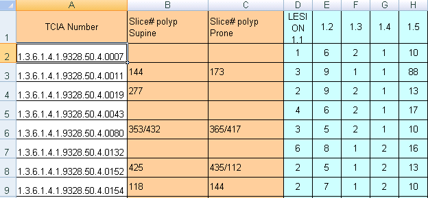
There are presently 825 cases in this collection with XLS sheets that provide polyp descriptions and their location within the colon segments. To link the XLS polyp tables with the DICOM image studies in TCIA you should understand that some cases in the TCIA are identified by long numbers with the last 4 digits after the last decimal point (e.g.: NCIA study number "1.3.6.1.4.1.9328.50.4.0040" referred to as case "40"). In addition there are a fewer number of additional positive cases that begin their identification number with 'CTC' (e.g.: CTC-5401799343)
Three related XLS spreadsheets are in this release.
- TCIA CTC large 10 mm polyps.xls - Contains the case numbers for 35 cases (out of the 825 total TCIA cases) where at least one 10mm or larger size polyp was found. Individual cases may have several (up to 20) polyps of different sizes listed on a particular XLS row as "LESION 1.x, 2.x,3.x etc. – see "feature key" below).
- TCIA CTC 6 to 9 mm polyps.xls - Contains 69 cases with smaller size polyps.
- TCIA CTC no polyp found.xls - Contains 243 cases that were recorded as free of polyps by both CTC and optical techniques.
Thus in this CT Colonography collection you will be able to download the prone and supine DICOM images from OC same-day validated 243 negative cases, 69 cases with 6 to 9 mm polyps, and 35 cases which have at least one > 10 mm polyp and their histological type. Below is the key for deciphering the features in the spreadsheet.
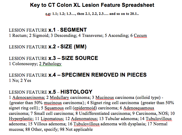
WARNING: NCI cannot assure archive users of error-free validity of the XL polyp location data since NCI did not itself perform the clinical study or its analysis.
You will note that two XLS files with positive findings have multiple columns descriptors of individual polyp lesions listed as in the table below. The meaning of the colored columns labeled "LESION 1.1...1.2...1.3...1.4, etc" is explained in the attached key-code ".tiff" file entitled "Polyp description key table.tiff"). Some CT scan slice numbers where the polyps were found are provided, but unfortunately the table may not have complete slice number information – you'll just have to do the best you can with the data NCI was given.
Citations & Data Usage Policy
This collection is freely available to browse, download, and use for commercial, scientific and educational purposes as outlined in the Creative Commons Attribution 3.0 Unported License. See TCIA's Data Usage Policies and Restrictions for additional details. Questions may be directed to help@cancerimagingarchive.net. Please be sure to acknowledge both this data set and TCIA in publications by including the following citations in your work:
CT Colonography Citation
The Cancer Imaging Archive Team. Data From CT_Colonography. doi:10.7937/K9/TCIA.2015.NWTESAY1
TCIA Citation
Clark K, Vendt B, Smith K, Freymann J, Kirby J, Koppel P, Moore S, Phillips S, Maffitt D, Pringle M, Tarbox L, Prior F. The Cancer Imaging Archive (TCIA): Maintaining and Operating a Public Information Repository, Journal of Digital Imaging, Volume 26, Number 6, December, 2013, pp 1045-1057. (paper)
Other Publications Using This Data
- NEJM publication for their published results (N Engl J Med. 2008 Sep 18;359(12):1207-17)
- Also see Publications#Collection:CTColonography
If you have a manuscript you'd like to add please contact the TCIA Helpdesk.
Version 1 (Current): Updated 2013/11/15
| Data Type | Download all or Query/Filter |
|---|---|
| Images (DICOM, 462.6GB) | |
| Polyp Descriptions - Large 10mm (XLS) | |
| Polyp Descriptions - 6 to 9mm (XLS) | |
| Polyp Descriptions - No polyp found (XLS) |
Data Access
Imaging Data
You can view and download these images on The Cancer Imaging Archive by clicking and selecting the Breast-Diagnosis collection.
Collection Statistics |
|
|---|---|
Modalities | MR (with some PET/CT) |
Number of Patients | 88 |
Number of Studies | 148 |
Number of Series | 429 |
Number of Images | 105,050 |
| Image Size (GB) | 1.16 |
If you are unsure how to download this Collection please view our quick guide on Searching by Collection or refer to our The Cancer Imaging Archive User's Guide for more detailed instructions on using the site.
Metadata
This collection includes a spreadsheet (updated 7/16/11) with BIRADS MRI features from the imaging report and denoted key image slice with the approximate X-Y center position if a mass was found. Key clinical features and abstracts of the pathology report including ER, PR and HER2 results and Oncotype score are included when available.