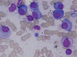
Summary
Additional Notes
This collection has also been uploaded to the Harvard Blood Cancer Dataverse website. Please refer to DOI 10.7910/DVN/XCX7ST for more information.
Additional Publications using this dataset:
- Ritu Gupta, Pramit Mallick, Rahul Duggal, Anubha Gupta, and Ojaswa Sharma, "Stain Color Normalization and Segmentation of Plasma Cells in Microscopic Images as a Prelude to Development of Computer Assisted Automated Disease Diagnostic Tool in Multiple Myeloma," 16th International Myeloma Workshop (IMW), India, March 2017 https://doi.org/10.1016/j.clml.2017.03.178
Data Access
| Data Type | Download all or Query/Filter | License |
|---|---|---|
| Slide Images (BMP, 1.27GB) | (Download and apply the IBM-Aspera-Connect plugin to your browser to retrieve this faspex package) | |
| Annotated plasma cell images (PDF, 12.67 MB) |
|
Detailed Description
Image Statistics | Pathology Imaging Statistics |
|---|---|
Modalities | Pathology |
Number of Participants | 5 |
Number of Studies | 5 |
Number of Images | 85 |
| Images Size (GB) | 1.27 |
Citations & Data Usage Policy
Users must abide by the TCIA Data Usage Policy and Restrictions. Attribution should include references to the following citations:
Data Citation
Gupta, R., & Gupta, A. (2019). MiMM_SBILab Dataset: Microscopic Images of Multiple Myeloma [Data set]. The Cancer Imaging Archive. https://doi.org/10.7937/tcia.2019.pnn6aypl
Publication Citation
Gupta, A., Duggal, R., Gehlot, S., Gupta, R., Mangal, A., Kumar, L., Thakkar, N., & Satpathy, D. (2020). GCTI-SN: Geometry-inspired chemical and tissue invariant stain normalization of microscopic medical images. Medical Image Analysis, 65, 101788. https://doi.org/10.1016/j.media.2020.101788
Publication Citation
Gupta, A., Mallick, P., Sharma, O., Gupta, R., & Duggal, R. (2018). PCSeg: Color model driven probabilistic multiphase level set based tool for plasma cell segmentation in multiple myeloma. PLOS ONE, 13(12), e0207908. https://doi.org/10.1371/journal.pone.0207908
TCIA Citation
Clark, K., Vendt, B., Smith, K., Freymann, J., Kirby, J., Koppel, P., Moore, S., Phillips, S., Maffitt, D., Pringle, M., Tarbox, L., & Prior, F. (2013). The Cancer Imaging Archive (TCIA): Maintaining and Operating a Public Information Repository. Journal of Digital Imaging, 26(6), 1045–1057. https://doi.org/10.1007/s10278-013-9622-7
Other Publications Using This Data
TCIA maintains a list of publications which leverage TCIA data. If you have a manuscript you'd like to add please contact TCIA's Helpdesk.
Version 1 (Current): Updated 2019/03/25
| Data Type | Download all or Query/Filter |
|---|---|
| Images (BMP, 1.27GB) | (Download and apply the IBM-Aspera-Connect plugin to your browser to retrieve this faspex package) |
| Annotated plasma cell images (PDF) |
