Summary
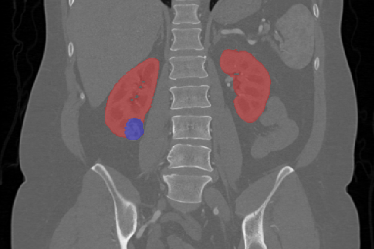
The imaging was collected during routine care of patients who were treated by either partial or radical nephrectomy at the University of Minnesota Medical Center. Many of the CT scans were acquired at referring institutions and are therefore heterogeneous in terms of scanner manufacturers and acquisition protocols. Semantic segmentations were performed by students under the supervision of an experienced urologic cancer surgeon.
Protocol
Please refer to the data descriptor manuscript for a comprehensive account of the data collection and annotation process - arXiv:1904.00445. The Clinical Trial Time Point is calculated from Day of Surgery.
Acknowledgements
We would like to acknowledge the following institutions for their support of the data collection and associated challenge:
- Climb 4 Kidney Cancer - Many of the students who worked on the chart review and manual segmentations for this dataset were graciously supported by Climb 4 Kidney Cancer (C4KC) as "C4KC Scholars"
- Intuitive Surgical - A prize of $5,000 was awarded by Intuitive Surgical to the KiTS19 Challenge's highest scoring team
- The National Cancer Institute of The National Institutes of Health - This work was supported under Award Number R01CA225435. The content is solely the responsibility of the authors and does not necessarily represent the official views of the National Institutes of Health
Harmonization of the components of this dataset, including into standard DICOM representation, was supported in part by the NCI Imaging Data Commons consortium. NCI Imaging Data Commons consortium is supported by the contract number 19X037Q from Leidos Biomedical Research under Task Order HHSN26100071 from NCI
Data Access
| Data Type | Download all or Query/Filter | License |
|---|---|---|
| Images and Segmentations (DICOM, 40.7GB) | (Download requires the NBIA Data Retriever) | |
| Clinical Data (CSV, 82 kB) |
Additional Resources for this Dataset
The NCI Cancer Research Data Commons (CRDC) provides access to additional data and a cloud-based data science infrastructure that connects data sets with analytics tools to allow users to share, integrate, analyze, and visualize cancer research data.
- Imaging Data Commons (IDC) (Imaging Data)
Detailed Description
Collection Statistics | Radiology Image Statistics |
|---|---|
Modalities | CT, SEG |
Number of Participants | 210 |
Number of Studies | 210 |
Number of Series | 621 |
Number of Images | 71,423 |
| Image Size (GB) | 40.7 |
Note:
The segmentation corresponds to the arterial phase in every case. No processing or analysis was done on the other phases.
Citations & Data Usage Policy
Users must abide by the TCIA Data Usage Policy and Restrictions. Attribution should include references to the following citations:
Data Citation
Heller, N., Sathianathen, N., Kalapara, A., Walczak, E., Moore, K., Kaluzniak, H., Rosenberg, J., Blake, P., Rengel, Z., Oestreich, M., Dean, J., Tradewell, M., Shah, A., Tejpaul, R., Edgerton, Z., Peterson, M., Raza, S., Regmi, S., Papanikolopoulos, N., Weight, C. (2019) Data from C4KC-KiTS [Data set]. The Cancer Imaging Archive. DOI: 10.7937/TCIA.2019.IX49E8NX
Publication Citation
Heller, N., Isensee, F., Maier-Hein, K. H., Hou, X., Xie, C., Li, F., Nan, Y., Mu, G., Lin, Z., Han, M., Yao, G., Gao, Y., Zhang, Y., Wang, Y., Hou, F., Yang, J., Xiong, G., Tian, J., Zhong, C., Ma, J., Rickman, J., Dean, J., Stai, B., Tejpaul, R., Oestreich, M., Blake, P., Kaluzniak, H., Raza, S., Rosenberg, J., Moore, K., Walczak, E., Rengel, Z., Edgerton, Z., Vasdev, R., Peterson, M., McSweeney, S., Peterson, S., Kalapara, A., Sathianathen, N., Papanikolopoulos, N., Weight, C. (2021). The state of the art in kidney and kidney tumor segmentation in contrast-enhanced CT imaging: Results of the KiTS19 challenge. Medical Image Analysis, 67, 101821. https://doi.org/10.1016/j.media.2020.101821
TCIA Citation
Clark K, Vendt B, Smith K, Freymann J, Kirby J, Koppel P, Moore S, Phillips S, Maffitt D, Pringle M, Tarbox L, Prior F. (2013) The Cancer Imaging Archive (TCIA): Maintaining and Operating a Public Information Repository, Journal of Digital Imaging, Volume 26, Number 6, December, 2013, pp 1045-1057. DOI: 10.1007/s10278-013-9622-7
Other Publications Using This Data
TCIA maintains a list of publications which leverage our data. If you have a publication you'd like to add please contact TCIA's Helpdesk.
Version 3 (Current): Updated 2020/06/18
| Data Type | Download all or Query/Filter |
|---|---|
Images and Segmentations (DICOM, 40.7GB) | (Requires NBIA Data Retriever) |
| Clinical Data (CSV, 82 kB) |
Upon initial publication of this dataset the segmentations were stored as sagittal series, while the CT images are axial. This caused difficulties loading this dataset into various DICOM tools. Those segmentations have now been converted (in a lossless fashion) to axial to resolve these issues.
Version 2: Updated 2020/03/23
| Data Type | Download all or Query/Filter |
|---|---|
Images and Segmentations (DICOM, 40.7GB) | |
| Clinical Data (CSV, 82 kB) |
Added clinical data spreadsheet.
Version 1: Updated 2019/12/18
| Data Type | Download all or Query/Filter |
|---|---|
Images (DICOM, 40.7GB) | Unavailable, see version 3 note. |