Summary
Investigators at the Mayo Clinic, with funding from the National Institute of Biomedical Imaging and Bioengineering (EB 017095 and EB 017185), have built a library of CT patient projection data in an open and vendor-neutral format. This format, referred to as DICOM-CT-PD (1), is an extended DICOM format that contains CT projection data and acquisition geometry. The de-identified patient projection data in the library were decoded with help of the manufacturer and have been converted into an open standardized format.
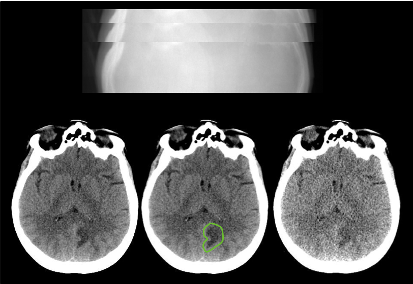
Reconstructed images, patient age and gender, and pathology annotation are also provided for these de-identified data sets. The library consists of scans from various exam types, including non-contrast head CT scans acquired for acute cognitive or motor deficit, low-dose non-contrast chest scans acquired to screen high-risk patients for pulmonary nodules, and contrast-enhanced CT scans of the abdomen acquired to look for metastatic liver lesions.
Acknowledgements
This work would not have been possible without the support and efforts of many individuals and organizations.
- A complete list of acknowledgements can be found here.
Data Access
Click the Download button to save a ".tcia" manifest file to your computer, which you must open with the NBIA Data Retriever. Click the Search button to open our Data Portal, where you can browse the data collection and/or download a subset of its contents.
| Data Type | Download all or Query/Filter |
|---|---|
| Images (DICOM, 1.23 TB) |
(Requires NBIA Data Retriever.) |
Images (DICOM, 2.0 GB) - Phantom Object Only |
|
| DICOM-CT-PD User Manual Version 3 (.pdf) |  |
Matlab DICOM-CTPD data dictionary (.txt) |
|
| Matlab DICOM-CTPD reader script (.txt) |
|
| Clinical Data (CSV, zip) |
|


Click the Versions tab for more info about data releases.
Detailed Description
Image Statistics | |
|---|---|
Modalities | CT |
Number of Patients | 300 |
Number of Studies | 599 |
Number of Series | 1048 |
Number of Images | 13,027,273 |
| Images Size (TB) | 1.23 |
For each patient CT scan, three types of data are provided: DICOM-CT-PD projection data, DICOM image data, and Excel clinical data reports. CT projection data are provided for both full and simulated lower dose levels and CT image data reconstructed using the commercial CT system are provided for the full dose projection data. For patients scanned on the SOMATOM Definition Flash CT scanner from Siemens Healthcare, CT image data reconstructed using the commercial CT system are also provided for the lower dose projection data. All CT images were reconstructed using a filtered back projection method. Several instructional documents are provided to help users extract needed information from the DICOM-CT-PD files, including a dictionary file for the DICOM-CT-PD format, a DICOM-CT-PD reader, and a user manual.
This collection comprises 99 head scans (labeled N for neuro), 100 chest scans (labeled C for chest), and 100 abdomen scans (labeled L for liver). Fifty cases for each scan type are from a SOMATOM Definition Flash CT scanner (Siemens Healthcare, Forchheim, Germany). Forty-nine head cases, 50 chest cases, and 50 abdomen cases are from a Lightspeed VCT CT scanner (GE Healthcare, Waukesha, WI). Together, these data will greatly facilitate the development and validation of new CT reconstruction and/or denoising algorithms, including those associated with machine learning or artificial intelligence.
Acquisition protocol
All CT scans were acquired at routine dose levels for the practice at which they were obtained using standard-clinical protocols for the anatomical region of interest. Each clinical case was processed to include a second projection dataset at a simulated lower dose level. Head and abdomen cases are provided at 25% of the routine dose and chest cases are provided at 10% of the routine dose.
1Additional information regarding the CT projection data format: Chen B, Duan X, Yu Z, Leng S, Yu L, McCollough CH. Technical Note: Development and validation of an open data format for CT projection data. Med Phys. 2015;42(12):6964. (doi: https://doi.org/10.1118/1.4935406.)
Citations & Data Usage Policy
These collections are freely available to browse, download, and use for commercial, scientific and educational purposes as outlined in the Creative Commons Attribution 3.0 Unported License. See TCIA's Data Usage Policies and Restrictions for additional details. Questions may be directed to help@cancerimagingarchive.net.
Please be sure to acknowledge both this data set and TCIA in publications by including the following citations in your work:
Data Citation
McCollough, C.H., Chen, B., Holmes, D., III, Duan, X., Yu, Z., Yu, L., Leng, S., Fletcher, J. (2020). Data from Low Dose CT Image and Projection Data [Data set]. The Cancer Imaging Archive. https://doi.org/10.7937/9npb-2637
Grant Citation
Presentations and publications shall acknowledge grants EB017095 and EB017185 (Cynthia McCollough, PI) from the National Institute of Biomedical Imaging and Bioengineering.
TCIA Citation
Clark K, Vendt B, Smith K, Freymann J, Kirby J, Koppel P, Moore S, Phillips S, Maffitt D, Pringle M, Tarbox L, Prior F. The Cancer Imaging Archive (TCIA): Maintaining and Operating a Public Information Repository, Journal of Digital Imaging, Volume 26, Number 6, December, 2013, pp 1045-1057. DOI: 10.1007/s10278-013-9622-7
Other Publications Using This Data
TCIA maintains a list of publications which leverage TCIA data. If you have a manuscript you'd like to add please contact the TCIA Helpdesk.
Version 1 (Current): Updated yyyy/mm/dd
| Data Type | Download all or Query/Filter |
|---|---|
| Images (DICOM, 1.23 TB) | |
Images (DICOM, 2.0 GB) Phantom Object Only |
|
| DICOM-CT-PD User Manual Version 3 (.pdf) | |
Matlab DICOM-CTPD data dictionary (.txt) |
|
| Matlab DICOM-CTPD reader script (.txt) |
|
| Clinical Data (CSV, zip) |
|