Child pages
  • Decoding tumour phenotype by noninvasive imaging using a quantitative radiomics approach (Radiomics-Tumor-Phenotypes)

Versions Compared

Key

  • This line was added.
  • This line was removed.
  • Formatting was changed.

...

This data applies a radiomic approach to computed tomography data of 1,019 patients with lung or head-and-neck cancer which are described in Nature Communications (http://doi.org/10.1038/ncomms5006).  The various arms of the study are represented in TCIA as distinct Collections including NSCLC-Radiomics (Lung1), NSCLC-Radiomics-Genomics (Lung3), Head-Neck-Radiomics-HN1 (H&N1)NSCLC-Radiomics-Interobserver1 (Multiple delineation), and RIDER Lung CT Segmentation Labels from: Decoding tumour phenotype by noninvasive imaging using a quantitative radiomics approach (RIDER-LungCT-Seg) (RIDER test/retest).

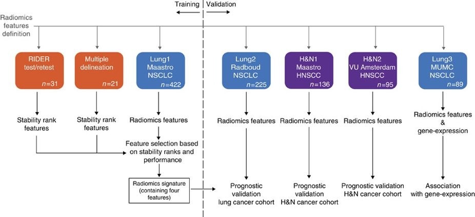
Radiomics refers to the comprehensive quantification of tumour phenotypes by applying a large number of quantitative image features. In present analysis 440 features quantifying tumour image intensity, shape and texture, were extracted. We found that a large number of radiomic features have prognostic power in independent data sets, many of which were not identified as significant before. Radiogenomics analysis revealed that a prognostic radiomic signature, capturing intra-tumour heterogeneity, was associated with underlying gene-expression patterns. These data suggest that radiomics identifies a general prognostic phenotype existing in both lung and head-and-neck cancer. This may have a clinical impact as imaging is routinely used in clinical practice, providing an unprecedented opportunity to improve decision-support in cancer treatment at low cost.

...

Localtab Group


Localtab
activetrue
titleData Access

Data Access

Click the Download button to save a ".tcia" manifest file to your computer, which you must open with the NBIA Data Retriever


Data TypeDownload all or Query/Filter
Image Data (DICOM) and Clinical Data

Please refer to each Collection page to download available images and clinical data:

Lung CT Segmentation Labels from: Decoding tumour phenotype by noninvasive imaging using a quantitative radiomics approach

NSCLC-Radiomics-Genomics (Lung3)

Gene Expression Data


Tcia button generator
labelSearch
urlhttp://www.ncbi.nlm.nih.gov/geo/query/acc.cgi?acc=GSE58661 
http://www.ncbi.nlm.nih.gov/geo/query/acc.cgi?acc=GSE58661 


Please contact help@cancerimagingarchive.net  with any questions regarding usage.


Collections Used in this Third Party Analysis

Below is a list of the Collections used in these analyses:

  • NSCLC-Radiomics (Lung1)
  • NSCLC-Radiomics-Genomics (Lung3)
  • Head-Neck-Radiomics-HN1 (H&N1)
  • Some data in this collection contains images that could potentially be used to reconstruct a human face. To safeguard the privacy of participants, users must sign and submit a TCIA No Commercial Limited Access License to help@cancerimagingarchive.net before accessing this portion of the data.

    RIDER Lung CT Segmentation Labels from: Decoding tumour phenotype by noninvasive imaging using a quantitative radiomics approach 

    Source Data Type

    Download

    License

    Corresponding Original Images from Head-Neck-Radiomics-HN1 (H&N1) (DICOM)


    Tcia button generator
    urlhttps://wiki.cancerimagingarchive.net/download/attachments/52762760/Head-Neck-Radiomics-HN1-Version%202-Sept%202019%20NBIA-manifest.tcia?version=1&modificationDate=1568995984096&api=v2


    Tcia button generator
    labelSearch
    urlhttps://nbia.cancerimagingarchive.net/nbia-search/?MinNumberOfStudiesCriteria=1&CollectionCriteria=HEAD-NECK-RADIOMICS-HN1


    (Download requires NBIA Data Retriever)

    Corresponding Original Images from NSCLC-Radiomics (Lung1), NSCLC-Radiomics-Genomics (Lung3), NSCLC-Radiomics-Interobserver1 (Multiple delineation)

     (DICOM)


    Tcia button generator
    urlhttps://wiki.cancerimagingarchive.net/display/DOI/Decoding+tumour+phenotype+by+noninvasive+imaging+using+a+quantitative+radiomics+approach?preview=%2F18514090%2F157287803%2Fmanifest-20230519_CC3-NC.tcia


    Tcia button generator
    labelSearch
    urlhttps://nbia.cancerimagingarchive.net/nbia-search/?MinNumberOfStudiesCriteria=1&CollectionCriteria=NSCLC-Radiomics,NSCLC-Radiomics-Interobserver1,NSCLC-Radiomics-Genomics


    (Download requires NBIA Data Retriever)

    Tcia cc by 3 nc

    Corresponding Original Images from RIDER-LungCT-Seg

    (RIDER test/retest)  (DICOM)


    Tcia button generator
    urlhttps://wiki.cancerimagingarchive.net/download/attachments/46334165/RIDER%20Lung%20CT%20RTSTRUCTS%20DICOM%20SEGS%20Leonard%20Wee%20Feb%2010%202020.tcia?api=v2


    (Download requires NBIA Data Retriever)


    Tcia cc by 3


    Localtab
    titleDetailed Description

    Detailed Description


    Localtab
    titleCitations & Data Usage Policy

    Citations & Data Usage Policy 

    Users of this data must abide by the Creative Commons Attribution-NonCommercial 3.0 Unported License under which it has been published. Questions may be directed to help@cancerimagingarchive.netAttribution should include references to the following citations:

    Tcia limited license policy

    Info
    titleDataset Citation

    Aerts, H., Velazquez, E. R., Leijenaar, R. T. H., Parmar, C., Grossmann, P., Carvalho, S., Bussink, J., Monshouwer, R., Haibe-Kains, B., Rietveld, D., Hoebers, F., Rietbergen, M. M., Leemans, C. R., Dekker, A., Quackenbush, J., Gillies, R. J., & Lambin, P. (2014). Data from: Decoding tumour phenotype by noninvasive imaging using a quantitative radiomics approach. [Data set]. The Cancer Imaging Archive. https://doi.org/10.7937/K9/TCIA.2014..UA0JGPDG

    In addition to the dataset citation above, please be sure to cite the following if you utilize these data in your research:


    Info
    titlePublication Citation

    Aerts, H. J. W. L., Velazquez, E. R., Leijenaar, R. T. H., Parmar, C., Grossmann, P., Carvalho, S., Bussink, J., Monshouwer, R., Haibe-Kains, B., Rietveld, D., Hoebers, F., Rietbergen, M. M., Leemans, C. R., Dekker, A., Quackenbush, J., Gillies, R. J., & Lambin, P. (2014). Decoding tumour phenotype by noninvasive imaging using a quantitative radiomics approach. Nature Communications, 5(1). https://doi.org/10.1038/ncomms5006


    Info
    titleTCIA Citation

    Clark, K., Vendt, B., Smith, K., Freymann, J., Kirby, J., Koppel, P., Moore, S., Phillips, S., Maffitt, D., Pringle, M., Tarbox, L., & Prior, F. (2013). The Cancer Imaging Archive (TCIA): Maintaining and Operating a Public Information Repository. Journal of Digital Imaging, 26(6), 1045–1057. https://doi.org/10.1007/s10278-013-9622-7


    Other Publications Using This Data

    TCIA maintains a list of publications that leverage TCIA our data. If you have a manuscript you'd like to add please contact the TCIA Helpdesk.


    Localtab
    titleVersions

    Version 2 (Current): 2020/03/23

    Added links to the recently published TCIA collections which reflect the additional arms of the study described in Nature Communications (http://doi.org/10.1038/ncomms5006).


    Data TypeDownload all or Query/Filter
    Image Data (DICOM) and Clinical Data

    Please refer to each Collection page to download available images and clinical data:

    NSCLC-Radiomics-Genomics (Lung3)

    Gene Expression Data

    Version 1

    (Current)

    : 2016/08/02

    Data TypeDownload all or Query/Filter
    Image Data (DICOM)

    Clinical Data (CSV, XLS)

    ClinicalMetadata

    Gene Expression Data


    ...