Description
This data applies a radiomic approach to computed tomography data of 1,019 patients with lung or head-and-neck cancer which are described in Nature Communications (http://doi.org/10.1038/ncomms5006). The various arms of the study are represented in TCIA as distinct Collections including NSCLC-Radiomics (Lung1), NSCLC-Radiomics-Genomics (Lung3), Head-Neck-Radiomics-HN1 (H&N1), NSCLC-Radiomics-Interobserver1 (Multiple delineation), and RIDER Lung CT Segmentation Labels from: Decoding tumour phenotype by noninvasive imaging using a quantitative radiomics approach (RIDER-LungCT-Seg) (RIDER test/retest).
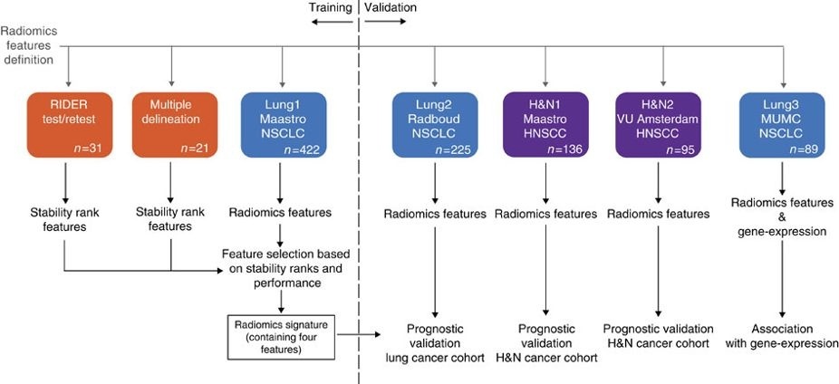
Radiomics refers to the comprehensive quantification of tumour phenotypes by applying a large number of quantitative image features. In present analysis 440 features quantifying tumour image intensity, shape and texture, were extracted. We found that a large number of radiomic features have prognostic power in independent data sets, many of which were not identified as significant before. Radiogenomics analysis revealed that a prognostic radiomic signature, capturing intra-tumour heterogeneity, was associated with underlying gene-expression patterns. These data suggest that radiomics identifies a general prognostic phenotype existing in both lung and head-and-neck cancer. This may have a clinical impact as imaging is routinely used in clinical practice, providing an unprecedented opportunity to improve decision-support in cancer treatment at low cost.
Data Access
| Data Type | Download all or Query/Filter |
|---|---|
| Image Data (DICOM) and Clinical Data | Please refer to each Collection page to download available images and clinical data:
|
Please contact help@cancerimagingarchive.net with any questions regarding usage.
Additional Resources for this Dataset
The following external resources have been made available by the data submitters. These are not hosted or supported by TCIA, but may be useful to researchers utilizing this collection.
- Genomics data in Gene Expression Omnibus for NSCLC-Radiomics-Genomics (Lung3) Gene Expression Data
Collections Used in this Analysis
Some data in this collection contains images that could potentially be used to reconstruct a human face. To safeguard the privacy of participants, users must sign and submit a TCIA No Commercial Limited Access License to help@cancerimagingarchive.net before accessing this portion of the data.
Source Data Type | Download | License |
|---|---|---|
Corresponding Original Images from Head-Neck-Radiomics-HN1 (H&N1) (DICOM) |
| |
Corresponding Original Images from NSCLC-Radiomics (Lung1), NSCLC-Radiomics-Genomics (Lung3), NSCLC-Radiomics-Interobserver1 (Multiple delineation) (DICOM) | (Download requires NBIA Data Retriever) | |
Corresponding Original Images from RIDER-LungCT-Seg (RIDER test/retest) (DICOM) | (Download requires NBIA Data Retriever) |
- NSCLC-Radiomics
- NSCLC-Radiomics-Genomics
- HEAD-NECK-RADIOMICS-HN1
- NSCLC-Radiomics-Interobserver1
- RIDER-LungCT-Seg
Detailed Description
Citations & Data Usage Policy
Users must abide by the TCIA Data Usage Policy and Restrictions. Attribution should include references to the following citations:
Data Citation
Aerts, H., Velazquez, E. R., Leijenaar, R. T. H., Parmar, C., Grossmann, P., Carvalho, S., Bussink, J., Monshouwer, R., Haibe-Kains, B., Rietveld, D., Hoebers, F., Rietbergen, M. M., Leemans, C. R., Dekker, A., Quackenbush, J., Gillies, R. J., & Lambin, P. (2014). Data from: Decoding tumour phenotype by noninvasive imaging using a quantitative radiomics approach (Radiomics-Tumor-Phenotypes). [Data set]. The Cancer Imaging Archive. https://doi.org/10.7937/K9/TCIA.2014..UA0JGPDG
Publication Citation
Aerts, H. J. W. L., Velazquez, E. R., Leijenaar, R. T. H., Parmar, C., Grossmann, P., Carvalho, S., Bussink, J., Monshouwer, R., Haibe-Kains, B., Rietveld, D., Hoebers, F., Rietbergen, M. M., Leemans, C. R., Dekker, A., Quackenbush, J., Gillies, R. J., & Lambin, P. (2014). Decoding tumour phenotype by noninvasive imaging using a quantitative radiomics approach. Nature Communications, 5(1). https://doi.org/10.1038/ncomms5006
TCIA Citation
Clark, K., Vendt, B., Smith, K., Freymann, J., Kirby, J., Koppel, P., Moore, S., Phillips, S., Maffitt, D., Pringle, M., Tarbox, L., & Prior, F. (2013). The Cancer Imaging Archive (TCIA): Maintaining and Operating a Public Information Repository. Journal of Digital Imaging, 26(6), 1045–1057. https://doi.org/10.1007/s10278-013-9622-7
Other Publications Using This Data
TCIA maintains a list of publications that leverage our data. If you have a manuscript you'd like to add please contact TCIA's Helpdesk.
Version 2 (Current): 2020/03/23
Added links to the recently published TCIA collections which reflect the additional arms of the study described in Nature Communications (http://doi.org/10.1038/ncomms5006).
| Data Type | Download all or Query/Filter |
|---|---|
| Image Data (DICOM) and Clinical Data | Please refer to each Collection page to download available images and clinical data:
|
NSCLC-Radiomics-Genomics (Lung3) Gene Expression Data |
Version 1 : 2016/08/02
| Data Type | Download all or Query/Filter |
|---|---|
| Image Data (DICOM) |
|
| Clinical Data (CSV, XLS) |
|
| Gene Expression Data |
|