Description
This data applies a radiomic approach to computed tomography data of 1,019 patients with lung or head-and-neck cancer which are described in Nature Communications (http://doi.org/10.1038/ncomms5006). The various arms of the study are represented in TCIA as distinct Collections including NSCLC-Radiomics (Lung1), NSCLC-Radiomics-Genomics (Lung3), Head-Neck-Radiomics-HN1 (H&N1), NSCLC-Radiomics-Interobserver1 (Multiple delineation), and RIDER Lung CT Segmentation Labels from: Decoding tumour phenotype by noninvasive imaging using a quantitative radiomics approach (RIDER-LungCT-Seg) (RIDER test/retest).
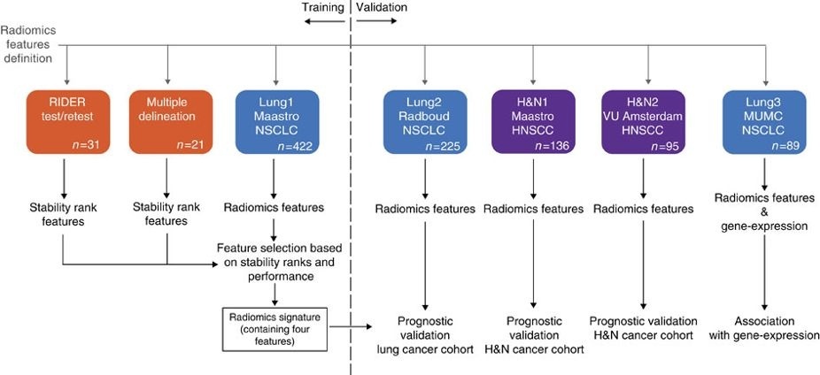
Radiomics refers to the comprehensive quantification of tumour phenotypes by applying a large number of quantitative image features. In present analysis 440 features quantifying tumour image intensity, shape and texture, were extracted. We found that a large number of radiomic features have prognostic power in independent data sets, many of which were not identified as significant before. Radiogenomics analysis revealed that a prognostic radiomic signature, capturing intra-tumour heterogeneity, was associated with underlying gene-expression patterns. These data suggest that radiomics identifies a general prognostic phenotype existing in both lung and head-and-neck cancer. This may have a clinical impact as imaging is routinely used in clinical practice, providing an unprecedented opportunity to improve decision-support in cancer treatment at low cost.
Data Access
| Data Type | Download all or Query/Filter |
|---|---|
| Image Data (DICOM) and Clinical Data | Please refer to each Collection page to download available images and clinical data:
|
NSCLC-Radiomics-Genomics (Lung3) Gene Expression Data |
Please contact help@cancerimagingarchive.net with any questions regarding usage.
Collections Used in this Third Party Analysis
Below is a list of the Collections used in these analyses:
Some data in this collection contains images that could potentially be used to reconstruct a human face. To safeguard the privacy of participants, users must sign and submit a TCIA No Commercial Limited Access License to help@cancerimagingarchive.net before accessing this portion of the data.
Source Data Type | Download | License |
|---|---|---|
Corresponding Original Images from Head-Neck-Radiomics-HN1 (H&N1) (DICOM) |
| |
Corresponding Original Images from NSCLC-Radiomics (Lung1), NSCLC-Radiomics-Genomics (Lung3), NSCLC-Radiomics-Interobserver1 (Multiple delineation) (DICOM) | (Download requires NBIA Data Retriever) | |
Corresponding Original Images from RIDER-LungCT-Seg (RIDER test/retest) (DICOM) | (Download requires NBIA Data Retriever) |
Detailed Description
Citations & Data Usage Policy
Users must abide by the TCIA Data Usage Policy and Restrictions. Attribution should include references to the following citations:
Dataset Citation
Aerts, H., Velazquez, E. R., Leijenaar, R. T. H., Parmar, C., Grossmann, P., Carvalho, S., Bussink, J., Monshouwer, R., Haibe-Kains, B., Rietveld, D., Hoebers, F., Rietbergen, M. M., Leemans, C. R., Dekker, A., Quackenbush, J., Gillies, R. J., & Lambin, P. (2014). Data from: Decoding tumour phenotype by noninvasive imaging using a quantitative radiomics approach. [Data set]. The Cancer Imaging Archive. https://doi.org/10.7937/K9/TCIA.2014..UA0JGPDG
Publication Citation
Aerts, H. J. W. L., Velazquez, E. R., Leijenaar, R. T. H., Parmar, C., Grossmann, P., Carvalho, S., Bussink, J., Monshouwer, R., Haibe-Kains, B., Rietveld, D., Hoebers, F., Rietbergen, M. M., Leemans, C. R., Dekker, A., Quackenbush, J., Gillies, R. J., & Lambin, P. (2014). Decoding tumour phenotype by noninvasive imaging using a quantitative radiomics approach. Nature Communications, 5(1). https://doi.org/10.1038/ncomms5006
TCIA Citation
Clark, K., Vendt, B., Smith, K., Freymann, J., Kirby, J., Koppel, P., Moore, S., Phillips, S., Maffitt, D., Pringle, M., Tarbox, L., & Prior, F. (2013). The Cancer Imaging Archive (TCIA): Maintaining and Operating a Public Information Repository. Journal of Digital Imaging, 26(6), 1045–1057. https://doi.org/10.1007/s10278-013-9622-7
Other Publications Using This Data
TCIA maintains a list of publications that leverage our data. If you have a manuscript you'd like to add please contact the TCIA Helpdesk.
Version 2 (Current): 2020/03/23
Added links to the recently published TCIA collections which reflect the additional arms of the study described in Nature Communications (http://doi.org/10.1038/ncomms5006).
| Data Type | Download all or Query/Filter |
|---|---|
| Image Data (DICOM) and Clinical Data | Please refer to each Collection page to download available images and clinical data:
|
NSCLC-Radiomics-Genomics (Lung3) Gene Expression Data |
Version 1 : 2016/08/02
| Data Type | Download all or Query/Filter |
|---|---|
| Image Data (DICOM) |
|
| Clinical Data (CSV, XLS) |
|
| Gene Expression Data |
|