Summary
| Redirect | ||||
|---|---|---|---|---|
|
| Excerpt |
|---|
| ACRIN 6657 was designed as a prospective study to test MRI for ability to predict response to treatment and risk-of-recurrence in patients with stage 2 or 3 breast cancer receiving neoadjuvant chemotherapy (NACT). ACRIN 6657 was conducted as a companion study to CALGB 150007, a correlative science study evaluating tissue-based biomarkers in the setting of neoadjuvant treatment of breast cancer. Collectively, CALGB 150007 and ACRIN 6657 formed the basis of the multicenter Investigation of Serial Studies to Predict Your Therapeutic Response with Imaging and moLecular Analysis (I-SPY TRIAL) breast cancer trial, a study of imaging and tissue-based biomarkers for predicting pathologic complete response (pCR) and recurrence-free survival (RFS). Additional information about the trial is available in the Study Protocol and Case Report Forms. |
Participant Eligibility and Enrollment: Criteria for inclusion were patients enrolling on CALGB 150007 with T3 tumors measuring at least 3 cm in diameter by clinical exam or imaging and receiving neoadjuvant chemotherapy with an anthracycline-cyclophosphamide regimen alone or followed by a taxane. Pregnant patients and those with ferromagnetic prostheses were excluded from the study. The study was open to enrollment from May 2002 to March 2006. 237 patients were enrolled, of which 230 met eligibility criteria.
Acknowledgements
This shared data set was provided by David Newitt, PhD and Nola Hylton, PhD from the Breast Imaging Research Program at UCSF, in collaboration with ACRIN, CALGB, the I-SPY TRIAL, and TCIA. Many thanks are due to The ACRIN 6657 trial team , The I-SPY 1 TRIAL team , and all the patients participating in these studies
Funding sources include NIH grants to UCSF (R01 CA132870 and U01 CA151235), ACRIN (UO1 CA079778 and UO1 CA080098), and CALGB (UO1 CA31964 and UO1 CA33601).
...
| Localtab Group | ||||||||
|---|---|---|---|---|---|---|---|---|
|
Choosing the Download option will provide you with a file to launch the TCIA Download Manager to download the entire collection. If you want to browse or filter the data to select only specific scans/studies please use the Search By Collection option.
|
...
|
...
Click the Versions tab for more info about data releases.
| Localtab | |||||||||||||||||||||||||||||||||||||||||||||||||||||||||||||||||||||||||||||||||||||
|---|---|---|---|---|---|---|---|---|---|---|---|---|---|---|---|---|---|---|---|---|---|---|---|---|---|---|---|---|---|---|---|---|---|---|---|---|---|---|---|---|---|---|---|---|---|---|---|---|---|---|---|---|---|---|---|---|---|---|---|---|---|---|---|---|---|---|---|---|---|---|---|---|---|---|---|---|---|---|---|---|---|---|---|---|---|
| |||||||||||||||||||||||||||||||||||||||||||||||||||||||||||||||||||||||||||||||||||||
Detailed Description
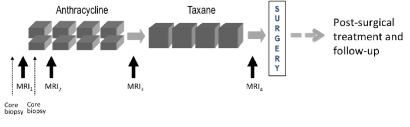
Requirements for MR imaging (As specified in the ACRIN 6657 protocol )Imaging time points: MRI exams were performed within four weeks prior to starting anthracycline-cyclophosphamide chemotherapy (T1, MRI1), at least 2 weeks after the first cycle of AC and prior to the second cycle of AC (T2, MRI2), between anthracycline-cyclophosphamide treatment and taxane therapy if taxane was administered (T3, MRI3), and after the final chemotherapy treatment and prior to surgery (T4, MRI4). The study schema is shown in Figure 1
Figure 1. CALGB 150007 and ACRIN 6657 study schema. Imaging protocol: MR imaging was performed on a 1.5 Tesla field strength scanner using a dedicated breast radiofrequency coil. The image acquisition protocol included a localization scan and T2-weighted sequence followed by a contrast-enhanced T1-weighted series. All imaging was performed unilaterally over the symptomatic breast and in the sagittal orientation. The contrast-enhanced series consisted of a high resolution (≤1mm in-plane spatial resolution) three-dimensional, fat-suppressed, T1-weighted gradient echo sequence with TR≤20 ms, TE = 4.5 ms, flip angle ≤ 45º, 16-18 cm field-of-view, minimum matrix 256x192, 64 slices, slice thickness ≤ 2.5 mm. Scan time length for the T1-weighted sequence was required to be between 4.5 and 5 minutes. The sequence was acquired once before contrast injection and repeated at least twice following injection. Tumor diameter measurement and volumetric analysis: Tumor longest diameter (LD) was measured by the site radiologist as the greatest extent of disease on baseline MR images, including intervening areas of non-enhancing tissue. The same measurement direction was used on all subsequent MRI exams. The primary predictor variable, functional tumor volume (FTV) was measured from contrast-enhanced images using the signal enhancement ratio (SER) method. Volumetric analysis, including Quality Control assessment, was performed centrally at the Breast Imaging Research Program (BIRP) laboratory at University of California at San Francisco (UCSF). Detailed information about the DICOM data is available in the DICOM Dictionary. Further information on these studies can be found at:
Imaging Data Transfer HistoryThe processing of the MR image data for ACRIN 6657 consisted of the following steps between image acquisition and the creation of this shared data set on TCIA:
While every effort was made to preserve the integrity of both the original image data and image meta-data (DICOM attributes, public and private), multiple file transfers and strict adherence to HIPPA guidelines for patient confidentiality may have resulted in loss of some data. If any questions arise, or patient PHI is found in any data on this collection, please contact the UCSF Breast Imaging Research Program (BIRP). Curated Data SetsIn addition to the complete set of ACRIN 6657 imaging studies ("Level 0" data), curated data sets based on UCSF QC assessment, protocol compliance and data completeness are provided for download in the form of TCIA shared lists. These include:
The image data sets are accompanied by Excel files with selected patient clinical and outcome data. Data Set DescriptionsLevel 0: Complete I-SPY 1 / ACRIN 6657 MRI DatasetThis data set is comprised of all HIPPA compliant, DICOM compliant MRI series. Level 0 image data set consists of 847 on-study MRI studies on 222 subjects in the UCSF image database. Level 1: MRI exams for which longest diameter was measuredThis data set is comprised of all studies with MRI measured longest diameter (LD) values reported.
Level 2a: Good SER Volume Dataset – updated 9/3/14, 9/17/16This data set is comprised of the patient studies which, following quality reviews in 2014 and 2016, were judged to have sufficiently good image quality and protocol compliance for volumetric DCE SER analysis. Rejection criteria included: incomplete volumetric DCE acquisitions, lack of a 2nd post-contrast acquisition, variability in fat suppression across the image, observed patient motion during the DCE acquisition, significant DCE protocol deviations such as changing scan parameters or image position during DCE acquisition. Level 2a image data set consists of 708 MR studies on 208 subjects in the UCSF image database. These include 7 studies not included in Level 1 (no MRI LD recorded) as listed in Table 2.
Level 2b: SER Volume Dataset Reported in Hylton et al. (Radiology, 2012) *This data set is comprised of the patient studies analyzed for pCR outcome and reported in the 2012 Radiology paper on ACRIN 6657 pCR results *. This data set is not provided as a shared list, as it is not recommended for use in further analysis. It is described here because it is the data set from which the Level 3 (primary aim analysis) set was derived. Inclusion and exclusion was determined by quality and protocol reviews available at that time. In addition to the exclusion criteria listed for Level 2a, studies done with imaging in the axial plane, in violation of the sagittal orientation specified in the trial imaging protocol, were excluded due to processing limitations of the analysis software. Similarly, bi-lateral sagittal acquisitions (alternating left and right volumetric acquisitions) were excluded. Level 2b image data set consists of 707 MRI studies on 207 subjects in the UCSF image database.
Tables 3 and 4 show the specific inclusion/exclusion differences between Levels 2a and 2b:
Level 3: Subset of Level 2b used in primary aim analysis, reported in Hylton et al. (Radiology, 2016) *This data set is comprised of the patient studies analyzed for RFS outcome and reported in the 2015 Radiology paper on ACRIN 6657 survival results (Hylton et al, Radiology *). Table 5 shows the 45 patients excluded from the level 2a cohort for this analysis. Please see the publication for specific information on exclusions of patients from this group. Level 3 image data set consists of 586 MRI studies on 162 subjects in the UCSF image database. This is also the study cohort used as the Test Phase data in the QIN BMMR Challenge.
Shared Lists For Curated Data SetsTo download image data sets for the curated data sets go to Tools / Search Shared Lists after logging in TCIA, and search for "ISPY1" Shared lists have been created for each of the main curated data sets: Level 1, 2a, and 3. For each level one may download either: "All Series" full MRI datasets including all original series and derived PE, SER maps and segmentation objects from the primary analysis "DCE + Derived" Original DCE series and derived PE, SER maps and segmentation objects from the primary analysis "DCE Only" Original DCE series only Relative sizes of the lists and full collection are shown in the Table below. Estimated download times can be found by loading the shared list into the download manager from the Search Shared Lists tool. Links are also provided in the Table for Excel spreadsheet files containing selected patient clinical data for the patients in each set. Further data will be made available in the future as it is released by I-SPY. To download: Tools / Search Shared Lists | |||||||||||||||||||||||||||||||||||||||||||||||||||||||||||||||||||||||||||||||||||||

| Localtab | ||||||||||
|---|---|---|---|---|---|---|---|---|---|---|
| ||||||||||
Citations & Data Usage PolicyThis collection is freely available to browse, download, and use for commercial, scientific and educational purposes as outlined in the Creative Commons Attribution 3.0 Unported License. See TCIA's Data Usage Policies and Restrictions for additional details. Questions may be directed to help@cancerimagingarchive.net. Please be sure to include the following citations in your work if you use this data set:
Other Publications Using This DataTCIA maintains a list of publications which leverage our data. At this time we are not aware of any publications based on this data. If you have a publication you'd like to add please contact the TCIA Helpdesk. |
| ||||||||||||||||||||||||||||||||||||||||||||||||||||||||||||||||||||||||||||||||||||||||||||||||||||||||||||||||||||||||||||||||||||||||||||||||||||||||||||||||||||||||||||||||||||||||||||||||||||||||||||||||||||||||||||||||||||||||||||||||||||||||||||||||||||||||||||||||||||||||
...
| title | Versions |
|---|
...
|
...
|
...