| Redirect | ||||
|---|---|---|---|---|
|
Summary
| Excerpt |
|---|
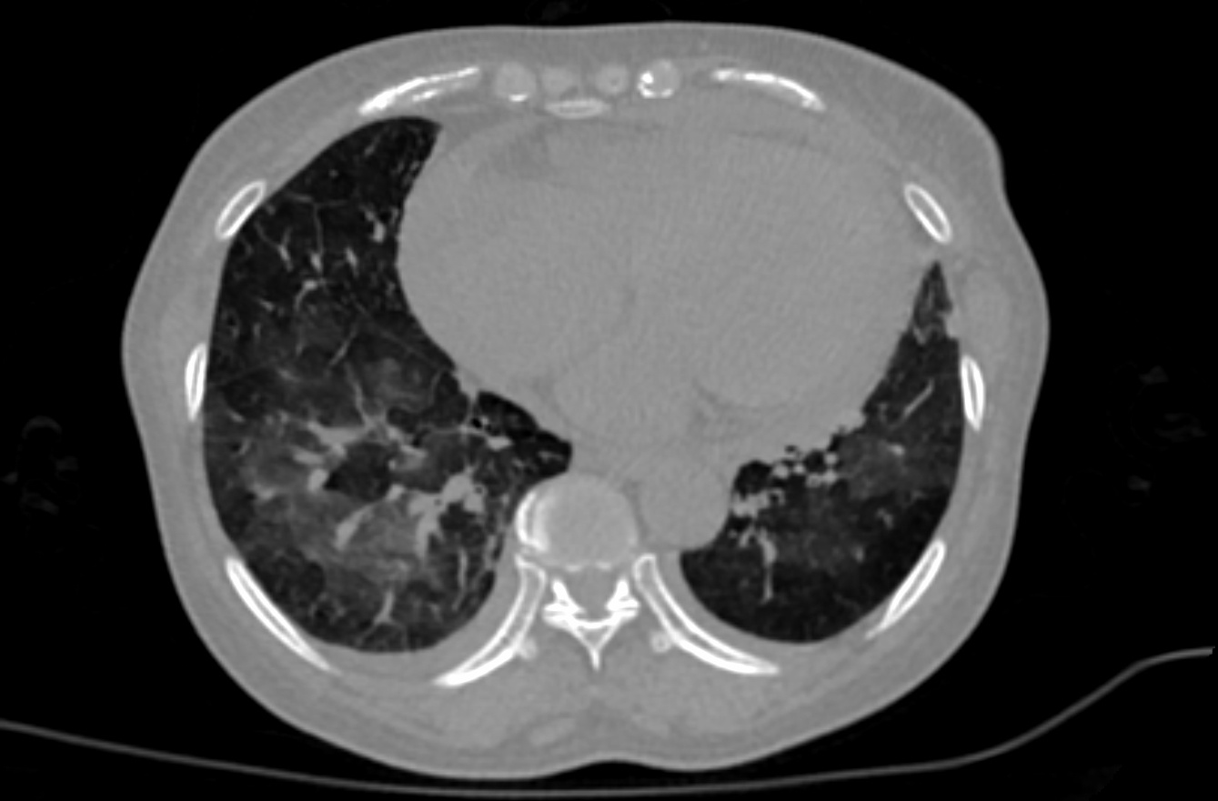
These retrospective NIfTI image datasets consists of unenhanced chest CTs:
The initial images for both datasets were acquired at the point of care in an outbreak setting from patients with Reverse Transcription Polymerase Chain Reaction (RT-PCR) confirmation for the presence of SARS-CoV-2. Patients presented to a health care setting with a combination of symptoms, exposure to an infected patient, or travel history to an outbreak region. All patients had a positive RT-PCR for SARS-CoV-2 from a sample obtained within 1 day of the initial CT. CT exams were performed without intravenous contrast and with a soft tissue reconstruction algorithm. The DICOM images were subsequently converted into NIfTI format. The second dataset also had other follow up CTs, in addition to the initial point of care CT. A multidisciplinary team trained several models using portions of the first dataset, along with additional CTs and manually annotated images from other sources. A classification model derived in part from the first dataset is described in a Nature Communications manuscript at: https://doi.org/10.1038/s41467-020-17971-2. The NVIDIA-related frameworks and models specific to this publication are available at no cost as part of the NVIDIA Clara Train SDK at https://ngc.nvidia.com/catalog/containers/nvidia:clara:ai-covid-19. This includes both inference-based pipelines for evaluation, as well as model weights for further training or fine tuning in outside institutions. The second data set of 121 serial / sequential CTs in 29 patients is reported in a Scientific Reports manuscript at https://doi.org/10.1038/s41598-021-85694-5. |
...

