Summary
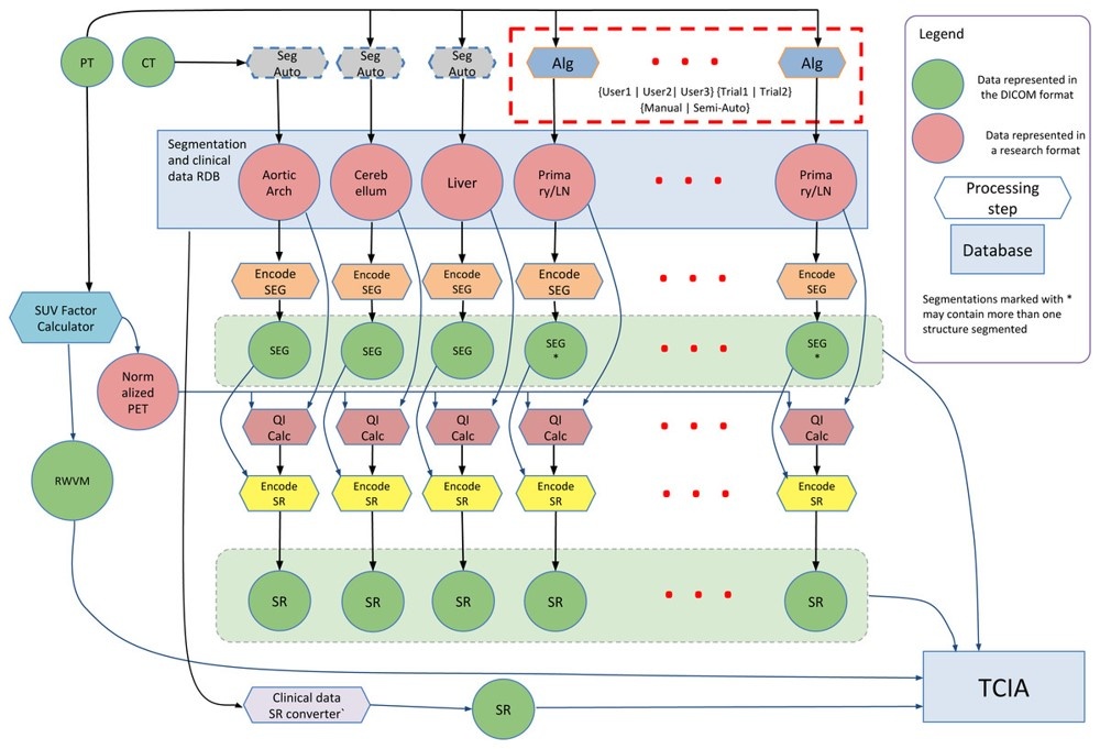
This collection is a set of head and neck cancer patients' multiple positron emission tomography/computed tomography (PET/CT) 18F-FDG scans–before and after therapy–with follow up scans where clinically indicated. The data was provided to help facilitate research activities of the National Cancer Institute's (NCI's) Quantitative Imaging Network (QIN). This collection was supported by Grants: U24 CA180918 (http://qiicr.org) and U01 CA140206.The following schematic summarizes much of the work done within the QIICR grant to augment the PET/CT scans with segmentations and clinical data using the DICOM standard: (click to enlarge)
About the NCI QIN
The mission of the QIN is to improve the role of quantitative imaging for clinical decision making in oncology by developing and validating data acquisition, analysis methods, and tools to tailor treatment for individual patients and predict or monitor the response to drug or radiation therapy. More information is available on the Quantitative Imaging Network Collections page. Interested investigators can apply to the QIN at: Quantitative Imaging for Evaluation of Responses to Cancer Therapies (U01) PAR-11-150.
Data Access
Click the Download button to save a ".tcia" manifest file to your computer, which you must open with the NBIA Data Retriever . Click the Search button to open our Data Portal, where you can browse the data collection and/or download a subset of its contents.
| Data Type | Download all or Query/Filter | License |
|---|---|---|
| Images and Segmentations (DICOM, 201.2 GB) | (Download requires the NBIA Data Retriever) | |
| Clinical Data (.xlsx 68 kB) | (See also Detailed Description tab) |
Click the Versions tab for more info about data releases.
Detailed Description
Collection Statistics | |
|---|---|
Modalities | PET, CT, SR, SEG, RWV |
Number of Participants | 279 |
Number of Studies | 1032 |
Number of Series | 3837 |
Number of Images | 701,002 |
| Image Size (GB) | 201.2 |
Associated Clinical Metadata
- Structured Report DICOM objects (Modality SR), are available for a subset of these subjects in the DICOM downloads and can be distinguished from image files by the series description "Clinical Data." Note, there is no image preview thumbnail for a Structured Report.
Citations & Data Usage Policy
Users must abide by the TCIA Data Usage Policy and Restrictions. Attribution should include references to the following citations:
Data Citation
Beichel R R, Ulrich E J, Bauer C, Wahle A, Brown B, Chang T, Plichta K A, Smith B J, Sunderland J J, Braun T, Fedorov A, Clunie D, Onken M, Magnotta VA, Menda Y, Riesmeier J, Pieper S, Kikinis R, Graham M M, Casavant T L, Sonka M, Buatti J M. (2015). Data From QIN-HEADNECK. The Cancer Imaging Archive. DOI: 10.7937/K9/TCIA.2015.K0F5CGLI
Publication Citation
Fedorov A, Clunie D, Ulrich E, Bauer C, Wahle A, Brown B, Onken M, Riesmeier J, Pieper S, Kikinis R, Buatti J, Beichel RR. (2016) DICOM for quantitative imaging biomarker development: a standards based approach to sharing clinical data and structured PET/CT analysis results in head and neck cancer research. PeerJ 4:e2057 DOI: 10.7717/peerj.2057
TCIA Citation
Clark K, Vendt B, Smith K, Freymann J, Kirby J, Koppel P, Moore S, Phillips S, Maffitt D, Pringle M, Tarbox L, Prior F. The Cancer Imaging Archive (TCIA): Maintaining and Operating a Public Information Repository, Journal of Digital Imaging, Volume 26, Number 6, December, 2013, pp 1045-1057. DOI: 10.1007/s10278-013-9622-7
Other Publications Using This Data
TCIA maintains a list of publications that leverage our data, including citations of this Collection. If you have a publication you'd like to add please contact the TCIA Helpdesk. Some publications that have used this dataset as a resource include
- Taghanaki et al. Segmentation-free direct tumor volume and metabolic activity estimation from PET scans. Comput Med Imaging Graph 2018 link to article
- Sinha et al. Towards automatic initialization of registration algorithms using simulated endoscopy images. link to article
- Ghattas, Andrew Emile Medical Imaging Segmentation Assessment via Bayesian Approaches to Fusion, Accuracy and Variability Estimation with Application to Head and Neck Cancer 2017 Thesis link
- Stoll et al. Comparison of Safety Margin Generation Concepts in Image Guided Radiotherapy to Account for Daily Head and Neck Pose Variations PLoS One 2016 link to article
- Ahmadvand et al. Tumor Lesion Segmentation from 3D PET Using a Machine Learning Driven Active Surface 2016 MLMI Conference Proceedings link to article
Version 4 (Current) : Updated 2020/09/15
| Data Type | Download all or Query/Filter |
|---|---|
| Images and Segmentations (DICOM 201.2 GB) | (Download requires the NBIA Data Retriever) |
| Clinical Data (.xlsx 68 kB) |
Added 123 new subjects (Patient IDs = QIN-HeadNeck-02-####). Added missing PT or CT pre-treatment and follow up scans to 28 of the previously existing QIN-HeadNeck-01-#### subjects. Added supporting clinical data in XLSX format for all patients.
Version 3: Updated 2019/07/24
| Data Type | Download all or Query/Filter |
|---|---|
| Images (DICOM, 103.5 GB) |
|
| DICOM Metadata Digest (CSV) |
Lifted restriction from SR object data download.
Version 2: Updated 2017/12/06
Downloads require the NBIA Data Retriever .
| Data Type | Download all or Query/Filter |
|---|---|
| Images (DICOM, 104 GB) | |
| DICOM Metadata Digest (CSV) |
Added associated DICOM SEG, SR, and RWV objects
Version 1: Updated 2015/08/20
| Data Type | Download all or Query/Filter |
|---|---|
| Images (DICOM, 102.76 GB) | |
| DICOM Metadata Digest (CSV) |