Summary
The National Institutes of Health Clinical Center performed 82 abdominal contrast enhanced 3D CT scans (~70 seconds after intravenous contrast injection in portal-venous) from 53 male and 27 female subjects. Seventeen of the subjects are healthy kidney donors scanned prior to nephrectomy. The remaining 65 patients were selected by a radiologist from patients who neither had major abdominal pathologies nor pancreatic cancer lesions. Subjects' ages range from 18 to 76 years with a mean age of 46.8 ± 16.7. The CT scans have resolutions of 512x512 pixels with varying pixel sizes and slice thickness between 1.5 − 2.5 mm, acquired on Philips and Siemens MDCT scanners (120 kVp tube voltage).
A medical student manually performed slice-by-slice segmentations of the pancreas as ground-truth and these were verified/modified by an experienced radiologist.
Data Access
Choosing the Download option will provide you with a file to launch the TCIA Download Manager to download the entire collection. If you want to browse or filter the data to select only specific scans/studies please use the Search By Collection option.
| Data Type | Download all or Query/Filter |
|---|---|
| Images (DICOM, 10.2 GB) |
|
| Manual Annotations |  |

Click the Versions tab for more info about data releases.
Detailed Description
Collection Statistics |
|
|---|---|
Modalities | CT |
Number of Patients | 82 |
Number of Studies | 82 |
Number of Series | 82 |
Number of Images | 19,328 |
| Image Size (GB) | 10.2 |
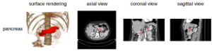
Data Example
Note
The DICOM files were created from anonymized volumetric images (Analyze and NifTI) using this from ITK: http://www.itk.org/Doxygen/html/Examples_2IO_2ImageReadDicomSeriesWrite_8cxx-example.html.
Citations & Data Usage Policy
This collection is freely available to browse, download, and use for commercial, scientific and educational purposes as outlined in the Creative Commons Attribution 3.0 Unported License. See TCIA's Data Usage Policies and Restrictions for additional details. Questions may be directed to help@cancerimagingarchive.net.
Please be sure to include the following citations in your work if you use this data set:
Data Citation
Holger R. Roth, Amal Farag, Evrim B. Turkbey, Le Lu, Jiamin Liu, and Ronald M. Summers. (2016). Data From Pancreas-CT. The Cancer Imaging Archive. http://doi.org/10.7937/K9/TCIA.2016.tNB1kqBU
Publication Citation
Roth HR, Lu L, Farag A, Shin H-C, Liu J, Turkbey EB, Summers RM. DeepOrgan: Multi-level Deep Convolutional Networks for Automated Pancreas Segmentation. N. Navab et al. (Eds.): MICCAI 2015, Part I, LNCS 9349, pp. 556–564, 2015. (paper)
TCIA Citation
Clark K, Vendt B, Smith K, Freymann J, Kirby J, Koppel P, Moore S, Phillips S, Maffitt D, Pringle M, Tarbox L, Prior F. The Cancer Imaging Archive (TCIA): Maintaining and Operating a Public Information Repository, Journal of Digital Imaging, Volume 26, Number 6, December, 2013, pp 1045-1057. (paper)
Other Publications Using This Data
If you have a publication you'd like to add please contact the TCIA Helpdesk.